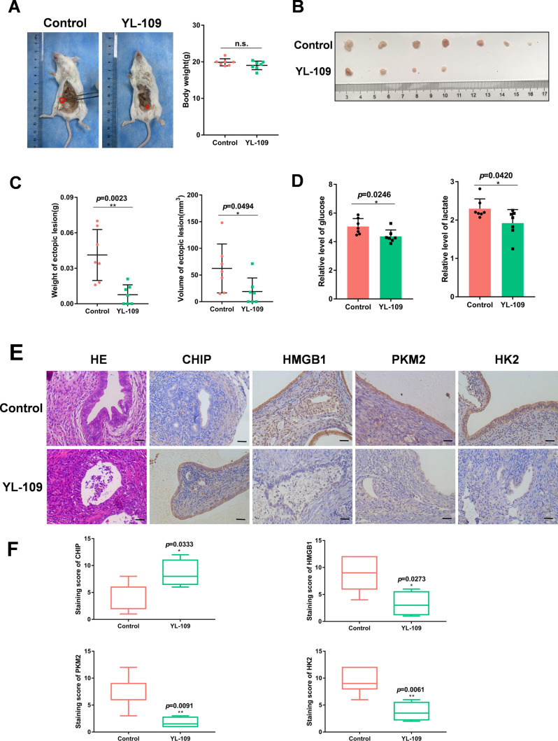
Fig. 3.
YL-109 inhibits the development of endometriosis. A–D After establishing the endometriosis mouse model, the control group was given intraperitoneal saline and the experimental group was given intraperitoneal YL-109 (15 mg/kg). After four weeks, the mice were executed to observe the growth and weight of ectopic tissues and detect the glucose and lactate levels in the peritoneal fluid. A The red circle represented the implanted ectopic tissue. B Represented the largest ectopic tissue of each mouse. E Representative photographs of H/E staining and CHIP, HMGB1, PKM2, HK2 staining of ectopic samples (scale bar, 20 µm). F Box plots showing the difference in immunoreactivity of CHIP, HMGB1, PKM2, HK2 in two groups. (All data represent mean ± SEM. The Student’s t test was used for data analysis. *P < 0.05, **P < 0.01, ***P < 0.001, ****P < 0.0001)

