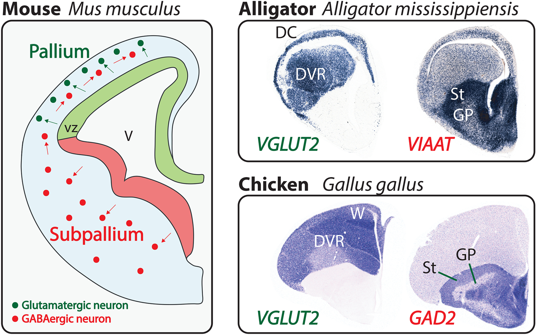
Figure 1.

The vertebrate pallium and subpallium.
Left: developing mouse telencephalon at 12.5 days of gestation, shortly after the onset of neocortical neurogenesis. One telencephalic hemisphere is diagrammed in cross-section with medial to the right and dorsal at the top. Neural progenitor cells line the ventricle (V), forming the ventricular zone (vz). Pallial progenitor cells (light green) give rise to excitatory, glutamatergic neurons (dark green), which migrate from their place of birth (green arrows) but remain within the pallium. In contrast, subpallial progenitor cells (light red) produce inhibitory, GABAergic neurons (dark red), which populate the subpallium but also disperse throughout the pallium (red arrows). This developmental pattern is conserved across the vertebrates. Top right: coronal sections through a late-embryonic alligator telencephalon labeled by in situ hybridization for VGLUT2 and VIAAT transcripts, which identify excitatory and inhibitory neurons, respectively [112]. Within the pallium, the three-layered dorsal cortex (DC) and the dorsal ventricular ridge (DVR) are identified. The striatum (St) and globus pallidus (GP) are two broadly conserved subdivisions of the subpallium. Bottom right: coronal sections through the telencephalon of a chicken hatchling labeled for VGLUT2 and GAD2 transcripts. Similar to non-avian reptiles, birds have a prominent DVR in the pallium. However, an additional nuclear structure, the Wulst (W), takes the place of a dorsal cortex. Note the GABAergic neurons scattered throughout the avian and reptilian pallia, as in mammals. Chicken sections from J. Rowell, Ragsdale laboratory.
