
Figure 4.
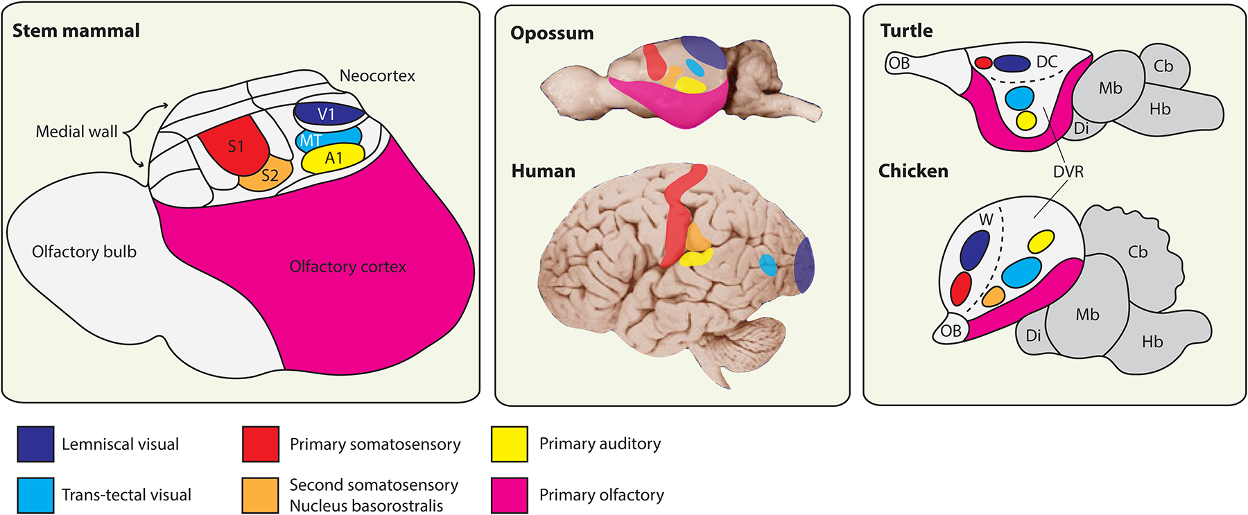
Evolution of telencephalic sensory centers in amniotes.
Left: reconstruction of telencephalon organization in a stem mammal based on a comparative analysis of neocortex in extant mammals, combined with information about brain proportions from early mammalian fossil skull endocasts (adapted from Kaas [95]). This mammal is inferred to have possessed a highly developed olfactory bulb and olfactory cortex, with a compact neocortex located dorsally. This small neocortex nonetheless contained a range of neocortical areas thought to be shared in all extant mammals, a subset of which are identified here (see [95] for further discussion). The primary visual area (V1) receives lemniscal visual input (which relays through the lateral geniculate nucleus of the dorsal thalamus), whereas the middle temporal visual area (MT) receives input from a separate, parallel visual pathway that relays through the optic tectum and then the thalamic lateral posterior nucleus. All mammals additionally share a primary auditory area (A1), a primary somatosensory area (S1) and an adjoining second somatosensory area (S2). Note that this nomenclature of cortical areas does not apply to birds and non-avian reptiles, which lack a neocortex. Middle: the sizes of neocortical sensory areas do not scale linearly with the overall surface area of the neocortical sheet. That is, mammals with a highly expanded neocortex, such as humans, have a larger proportion of non-primary-sensory and higher order association cortex. Differential allocation of cortical surface area is apparent, for example, in the large human frontal cortex rostral to S1. Placement of cortical areas based on [193], MT placement in opossum and human based on [197] and [44], respectively. Right: the core sensory pathways to the mammalian pallium are conserved also in birds and non-avian reptiles, where they target spatially discrete pallial domains. The lemniscal visual channel targets the dorsal cortex (DC) in turtles and the avian Wulst (W), whereas the trans-tectal visual channel targets defined nuclei deep within the dorsal ventricular ridge (DVR) in each species [106]. Primary somatosensory information targets the DC and the Wulst rostral to the lemniscal visual targets. Primary auditory information reaches the DVR in all known sauropsids. Birds possess an additional sensory nucleus in the rostral DVR, the nucleus basorostralis, which receives trigeminal somatosensory information via a direct projection from the hindbrain [6]. This nucleus expresses molecular markers of neocortical input neurons and is conserved in alligators, but possible homologies with mammalian features remain elusive [112]. Turtle and chicken schematics adapted from [114] and [120], respectively. Cb, cerebellum; Di, diencephalon; Hb, hindbrain; Mb, midbrain; OB, olfactory bulb.