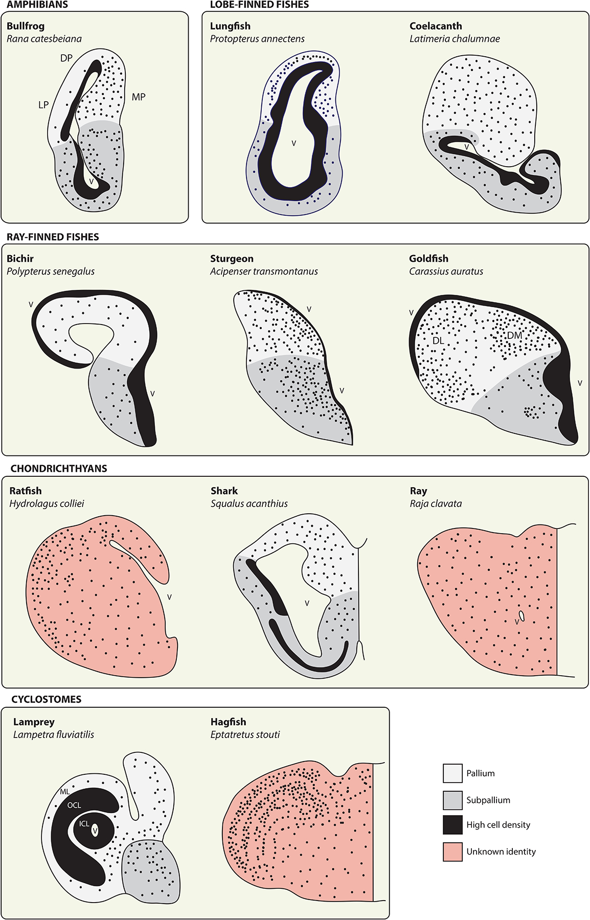
Figure 5.

The telencephala of anamniote vertebrates.
Schematics depict telencephalon anatomy from representatives of the anamniote vertebrates. Solid black territories represent regions of particularly high cell density, often along the surface of the lateral ventricle (V), whereas black dots provide a qualitative representation of cellular density and distribution. These illustrations are intended to provide a broad overview of telencephalon morphology in anamniotes and only the subset of neuroanatomical zones referred to in the main text are identified here. In all cases, pallial-subpallial boundaries should be regarded as approximations. See source materials for further details and discussion. Bullfrog adapted from [198] (DP, dorsal pallium; LP, lateral pallium; MP, medial pallium). Lungfish adapted from [24] and coelacanth from [199]. Ray-finned fishes adapted from [130] (DL, dorsolateral area; DM, dorsomedial area). Chondrichthyans adapted from [153]. Lamprey adapted from [175] (ICL, inner cellular layer; OCL, outer cellular layer; ML, molecular layer) and hagfish from [167].
