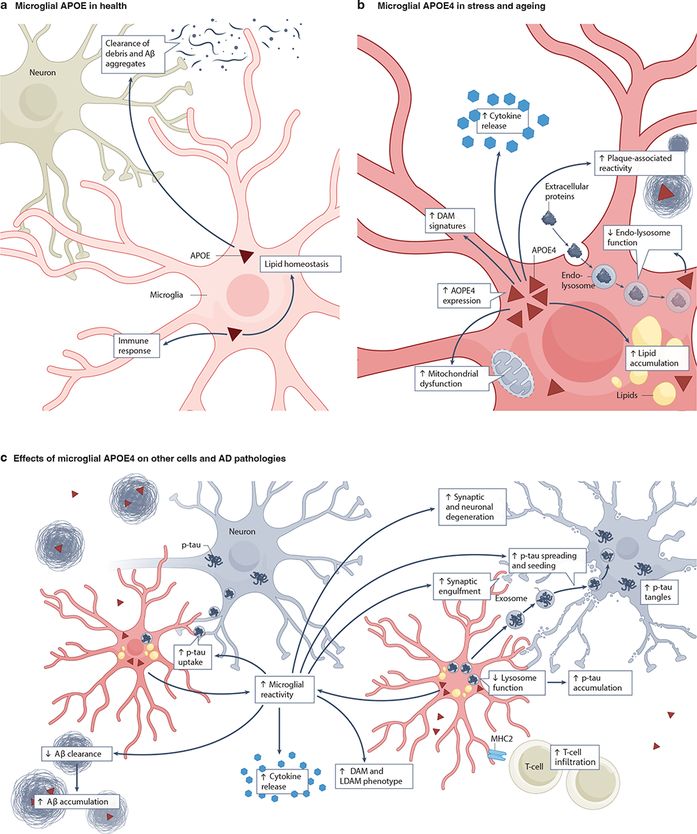
Fig. 4: Expression of APOE4 in microglia and its roles in AD pathogenesis.

a, Healthy microglia express low levels of apolipoprotein E (APOE)39. This APOE is involved in immune responses, lipid homeostasis and in clearing amyloid β (Aβ) aggregates and cell debris23,53,143. b, During stress and aging, microglia increase their APOE expression138–141. Microglial APOE4 leads to an increase in amyloid plaque-associated reactivity54, lipid accumulation27,148, lysosomal defects151,152, mitochondrial dysfunction141, disease-associated microglial (DAM) signatures140,167 and proinflammatory cytokine release167. c, Microglial APOE4 affects other cells, including neurons and T cells166, and contributes to AD pathogenesis by reducing Aβ clearance157 and lysosome function151 and by increasing synaptic engulfment161, p-tau uptake and accumulation86,151,152, DAM signatures140,152, lipid-droplet-accumulating microglia LDAM29,147, and cytokine release167. Together, these APOE4-induced microglial dysfunctions facilitate tau spreading and seeding162–164 and, ultimately, synaptic and neuronal degeneration (in conjunction with neuronal APOE4)140,152. The potential relationships among these microglial APOE4 effects on other cells and AD pathologies is depicted using arrows. Note that non-microglial sources of APOE have been omitted for clarity.
