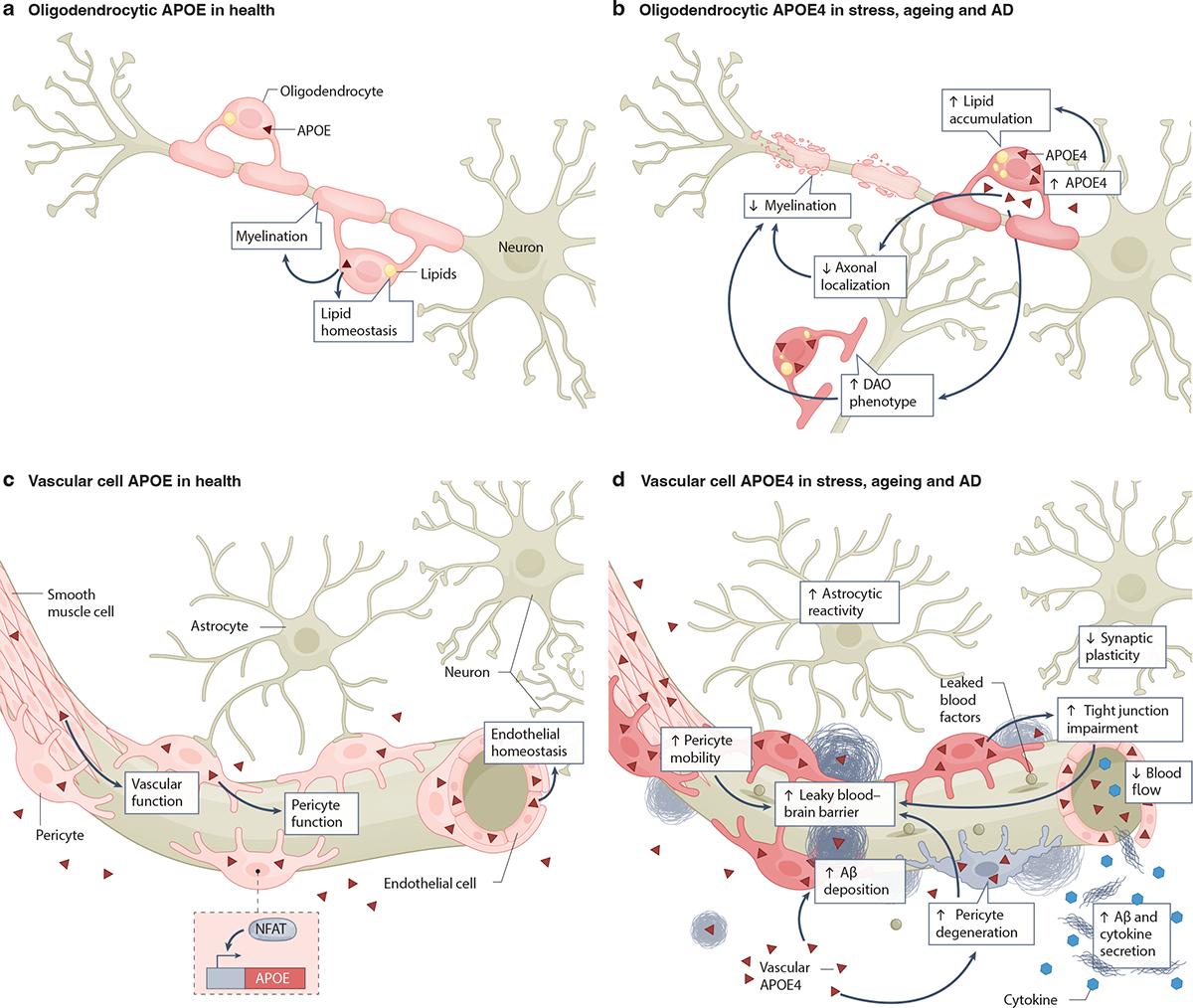
Fig. 5: Expression of APOE4 in oligodendrocytes and vascular cells and its roles in AD pathogenesis.

a, Healthy oligodendrocytes express low levels of apolipoprotein E (APOE)24,31,90. Oligodendrocytic APOE is involved in lipid homeostasis and myelination of neurons31. b, Under conditions of stress, aging, and AD, oligodendrocytes increase their APOE4 expression24,172. Oligodendrocytic APOE4 increases intracellular lipid accumulation and disease-associated oligodendrocyte (DAO) phenotypes24,31 and decreases oligodendrocytic axonal localization and myelination of neuronal axons31. The potential relationship among oligodendrocytic APOE4 effects on AD pathologies is depicted using arrows. c, Healthy cells of the brain vasculature, including smooth muscle cells, pericytes and endothelial cells, produce and secrete APOE39,176–182. The transcription factor NFAT regulates APOE expression in pericytes178. Vascular cell-produced APOE is involved in maintaining vascular function, pericyte function and endothelial homeostasis176–178,183. d, Under conditions of stress, aging, and AD, vascular cell APOE4 leads to tight junction impairments36,183, astrocyte reactivity185, increased pericyte migration and degeneration176,177, decreased synaptic plasticity185, reduced blood flow185, elevated secretion of amyloid β (Aβ) and cytokines180, increased Aβ deposition in the form of cerebral amyloid angiopathy (CAA)178, and blood-brain barrier (BBB) leakage35,36,176. The potential relationship among vascular APOE4 effects on other cells and AD pathologies is depicted using arrows. Note that non-oligodendrocytic and non-vascular sources of APOE4 have been omitted for clarity.
