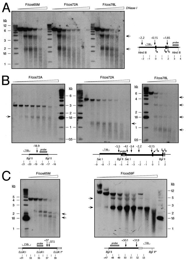
Figure 5.
DNase I-hypersensitive site mapping in cosmid transfectants. Nuclei from the indicated single-copy, intact cosmid transfectants were treated with increasing concentrations of DNase I. DNA was purified, digested with HindIII (A), BglII (B, left and right; C, right), SacI (B, center), or EcoRI (C, left) and analyzed by Southern hybridization using specific probes around α1AT described in Materials and Methods. The diagrams indicate the positions of relevant restriction sites, DHSs, probes and α1AT exons, using as coordinate zero an EcoRI site in the middle of exon IA of α1AT. Light gray boxes in (C) indicate a fragment of the SuperCos1 vector at the 3′ end of the transgene, and restriction sites with an asterisk are either in the vector or at the integration site of the cosmid in the rat genome. The ∼4.1 kb EcoRI fragment without DHSs (C, left; approximately position +31 to +35.1 kb) only hybridized weakly to the probe used, and is barely visible below the stronger ∼4.3 kb fragment recognized by the probe. Arrows beside the autoradiographs indicate the positions of sub-fragments generated by DNase I cleavage at specific DHSs. A 1 kb ladder was used as a size standard, and the positions of diagnostic bands are indicated beside the autoradiographs.

