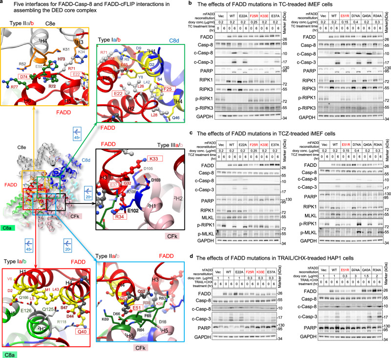
Fig. 6. Roles of the triple- and single-FADD models in hierarchical apoptotic signaling.
a Show five interfaces between FADDDED and tDED in the single-FADD structure. The interface residues of FADD are label in red, while FADD residues for mutagenesis are in red boxes. The side chains of the interface residues of FADD are shown as ball-and-sticks, while those of Casp-8 and cFLIP are shown as sticks. The interface residues of Casp-8 are colored as in Fig. 3a, while those of cFLIP are colored as in Supplementary Fig. 1a, respectively. The helices are numbered in Supplementary Fig. 2e. b Cell-based mutagenesis assays. FADD-reconstituted iMEF cells were treated with TNF/CHX (TC) to trigger apoptotic signaling. T TNFα, C CHX, Vec empty vecto, doxy doxycycline. F25R, K33E, and E51R mutants are highlighted in red because they are inactive in Supplementary Fig. 15c–f. c Same as b, but treated with TNF/CHX/zVAD (TCZ) to inhibit caspase activity and promote necroptotic signaling. Z zVAD. d Same as b, but FADD-reconstituted HAP1 cells were treated with TRAIL/CHX. All cell-based experiments were repeated twice with similar results. Uncropped blots are provided as a Source Data file.

