


Abstract
A recently described reaction for the UV-mediated attachment of alkenes to silicon surfaces is utilized as the basis for the preparation of functionalized silicon surfaces. UV light mediates the reaction of t-butyloxycarbonyl (t-BOC) protected ω-unsaturated aminoalkane (10-aminodec-1-ene) with hydrogen-terminated silicon (001). Removal of the t-BOC protecting group yields an aminodecane-modified silicon surface. The resultant amino groups can be coupled to thiol-modified oligodeoxyribonucleotides using a heterobifunctional crosslinker, permitting the preparation of DNA arrays. Two methods for controlling the surface density of oligodeoxyribonucleotides were explored: in the first, binary mixtures of 10-aminodec-1-ene and dodecene were utilized in the initial UV-mediated coupling reaction; a linear relationship was found between the mole fraction of aminodecene and the density of DNA hybridization sites. In the second, only a portion of the t-BOC protecting groups was removed from the surface by limiting the time allowed for the deprotection reaction. The oligodeoxyribonucleotide-modified surfaces were extremely stable and performed well in DNA hybridization assays. These surfaces provide an alternative to gold or glass for surface immobilization of oligonucleotides in DNA arrays as well as a route for the coupling of nucleic acid biomolecular recognition elements to semiconductor materials.
INTRODUCTION
Surfaces suitable for the immobilization of DNA have become an increasingly important biological tool in recent years. Arrays of DNA molecules, either as double-stranded segments or as short single-stranded oligodeoxyribonucleotides, have been utilized for drug development, DNA sequencing, medical diagnostics, nucleic acid–ligand binding studies and DNA computing (1–21). The principal advantages of using surface-bound oligonucleotides over those in solution include ease of purification, conservation of material and reagents, reduction of interference between oligonucleotides and facilitated sample handling (12). Previously explored surfaces for immobilization of DNA include latex beads (5), polystyrene (1), carbon electrodes (22–25), gold (17,23,26–28) and oxidized silicon or glass (3,8,11,29–32). The surface chemistries involved with these substrates do not generally possess all of the desired characteristics of an ideal surface. Such ideal surface characteristics include surface flatness and homogeneity, control of surface properties, thermal and chemical stability, reproducibility, and amenability to DNA immobilization and biochemical manipulation. More recently, the desire to use immobilized DNA as a biosensor in integrated circuits (33) would require the surface to be amenable to integration into a microelectronics format. The limitations of these previously used surfaces and attachment chemistries indicate the need to explore alternatives that more closely resemble the ideal. Unoxidized crystalline silicon offers advantages as a substrate for immobilization of DNA because of its high purity, highly organized and defined crystalline structure, robustness, and its ubiquitous use in the microelectronics industry.
Native silicon surfaces react with air under ambient conditions to form a thin surface layer of silicon oxide. This oxidized silicon surface is chemically similar to glass and suffers from some of the same drawbacks, namely inhomogeneity and variability in the relative number of Si–O–Si and Si–OH linkages. This inhomogeneity and the concomitant chemical variability it engenders can lead to difficulties in the reproducibility and homogeneity of the subsequent DNA-modified surfaces, particularly as the silane chemistry generally employed to couple to such surfaces is itself prone to stability problems and difficult-to-control polymerization processes (34,35). Recently, chemical pathways for direct functionalization of silicon substrates without an oxide layer has opened up new possibilities for highly controlled DNA attachment. These new attachment methods provide modified silicon surfaces through direct carbon–silicon bonds (19,36–41), and have resulted in methyl, chlorine, ester, or acid terminated substrates (36,39,40,42,43). Although Wagner used a N-hydroxysuccinimidyl ester terminated silicon surface to immobilize a 1752 bp double-stranded section of DNA (19), little other work of this nature has been reported. Recently, we have described a method to immobilize short oligonucleotides to a hydrogen-terminated silicon surface through an intervening chemical layer (polylysine) and heterobifunctional linker (43). This previous work demonstrated the successful immobilization and subsequent manipulation of short oligonucleotides on hydrogen-terminated silicon surfaces; however, as it includes the non-covalent association of a polymeric material to the surface it is necessarily less well-defined chemically than is the simpler and more direct surface attachment chemistry described here. The surface attachment chemistry described here does not rely on the non-covalent association of a polymer and permits control of the surface density of the DNA attachment sites, an important aspect of preparing reproducible and well-defined DNA arrays.
MATERIALS AND METHODS
All oligodeoxyribonucleotides were synthesized by the University of Wisconsin Biotechnology Center. Two types of oligonucleotides were employed for these experiments: oligonucleotides to be attached to the surface, and oligonucleotides employed for hybridization to the surface. Oligonucleotides to be attached to the surface were thiol-modified at either the 5′-end using the reagent 5′-Thiol-Modifier C6 (HSC6, Glen Research), or at the 3′-end using the reagent 3′-Thiol-Modifier C3 S-S CPG (C3SH, Glen Research); each of these oligonucleotides were 31 nt in length, comprised of a 15mer dT spacer sequence at the 5′-end, and a specific 16mer sequence at the 3′-end. The three 31mer sequences employed were 5′-HSC6-T15AA CGA TCG AGC TGC AA-3′ (S1), 5′-HSC6-T15AA CGA TGC AGG AGC AA-3′ (S2) and 5′-FAM-T15AA CGA TCG AGC TGC AA-C3SH-3′ (S3). In S3 ‘FAM’ refers to a fluorescein dye incorporated into the oligonucleotide during synthesis using the reagent 6-FAM phosphoramidite (Perkin-Elmer Biosystems). Oligonucleotides employed for hybridization to their surface-bound complements were 16 nt in length and complementary to the 3′ 16 nt of either S1 or S2. These oligonucleotides were also modified with FAM on the 5′-end. The thiol-modified oligonucleotides were deprotected according to guidelines from Glen Research (44,45) and purified by reversed-phase HPLC with a binary gradient elution. The 16 nt complements were purified in the same manner. Other reagents were obtained from Aldrich unless otherwise noted and are as follows: 1-dodecene, 10-undecenoyl chloride, sodium azide, trifluoroacetic acid (TFA), di-tert-butyl dicarbonate (Acros), sulfo-succinimidyl 4-(N-maleimidomethyl) cyclohexane-1-carboxylate (SSMCC) (Pierce), silicon (001) wafers (Virginia Semiconductor). Ultra pure water used for rinsing the wafers was obtained from a Millipore system.
Synthesis of 10-aminodec-1-ene
The 10-aminodec-1-ene used to modify the silicon surfaces was prepared using a variant of the Curtius reaction (46). A solution of 40.5 g of 10-undecenoyl chloride and tetrabutylammonium bromide (200 mg) was prepared in 300 ml of dichloromethane and cooled in an ice bath. Sodium azide (15.6 g) was dissolved in 50 ml of water and added to the mixture. This mixture was stirred continuously for 2 h in an ice bath. The organic layer was removed, washed twice with 50 ml of water, dried over anhydrous magnesium sulfate for 36 h and filtered. TFA (20.3 ml) was added slowly with stirring and the mixture was refluxed for 6 h. The resultant mixture containing the trifluoroacetamide was cooled and washed twice with 50 ml of saturated aqueous sodium bicarbonate, then dried over magnesium sulfate and filtered. The solvent was removed by rotary evaporation to yield an oil which was vacuum distilled to produce the pure trifluoroacetamide. NMR confirmed the identity of the trifluoroacetamide. Cleavage of the amide bond in a 10 ml portion of the trifluoroacetamide was accomplished by refluxing for 3 h in 500 ml of a 7% potassium carbonate methanol:water (2:5) solution (47). The free amine was extracted twice with 100 ml of ether, which was again dried over magnesium sulfate and filtered. Removal of the solvent by rotary evaporation and vacuum distillation yielded the product. NMR confirmed its identity as the pure amine.
Synthesis of the t-butyloxycarbonyl (t-BOC) protected amine
t-BOC protected 10-aminodec-1-ene was prepared by standard methods (48). A portion of the purified amine (5.11 g) was dissolved in 60 ml of chloroform that was added to a solution of 3 g NaHCO3 in 50 ml of water. Sodium chloride (6.45 g) was added along with 7.18 g of di-tert-butyl dicarbonate dissolved in a few milliliters of chloroform. This mixture was refluxed for 90 min and extracted twice with 50 ml of ether. The collected organic extracts were dried over magnesium sulfate, filtered and the ether removed by rotary evaporation. The t-BOC protected product was purified by vacuum distillation, and its identity was confirmed by NMR. Treating a 10 µl aliquot of this product with 1 ml of 25% TFA in methylene chloride for 1 h at room temperature allowed the free amine to be recovered which was verified by thin layer chromatography.
Reaction chamber
The chamber used for the UV mediated reaction consisted of a cylindrical aluminum base 11.5 cm in diameter and 5 cm tall. The interior of the base was milled to create a chamber 9 cm in diameter and 4 cm deep. A thin Teflon disk was placed in the bottom of this chamber. Inlet and outlet ports were added to allow for the continual passage of nitrogen gas. A Teflon lid 11.5 cm in diameter and 3.5 cm thick with an 8 cm diameter section removed from the middle was machined to fit over the aluminum base. The 8 cm diameter hole was milled to secure a quartz window. When assembled and in use the lid was secured and a UV lamp was placed over the chamber. The UV lamp was purchased from UV Products (part number CPQ-7446) and consisted of a low-pressure mercury vapor quartz grid lamp mounted on an aluminum reflector. The intensity of the lamp at 15 cm was 2000 µW/cm2 (254 nm).
Preparation of DNA-modified surface
Silicon (001) wafers were prepared for modification as previously described (19,43). The wafers were briefly exposed to a 2% solution of hydrofluoric acid in water to hydrogen terminate the surface atoms (note: hydrofluoric acid is extremely caustic; proper safety precautions should be followed). The wafers were then immediately placed in the reaction chamber, covered with 10–30 µl of the t-BOC protected 10-aminodec-1-ene, and exposed to UV light from the UV lamp for 2 h. The volume of the t-BOC protected 10-aminodec-1-ene used was enough to just cover the surface and varied with the size of the wafers. The thickness of the layer covering the wafer was not directly measured or controlled but was typically <1 mm. The modified surfaces were then subjected to 25% TFA in methylene chloride followed by a 3 min rinse in 10% NH4OH to remove the t-BOC protecting group and form the primary amine terminated surface. These surfaces were covered with 50 µl of a solution of the heterobifunctional crosslinker SSMCC (1.5 mM in 100 mM triethanolamine buffer, pH 7) for 20 min. SSMCC contains an amine reactive N-hydroxysuccinimide ester as well as a thiol-reactive maleimide moiety. The maleimide activated surfaces were reacted overnight with 0.4–0.8 µl droplets of ~1 mM thiol-modified DNA to form the DNA arrays. The DNA-modified surfaces were then rinsed with distilled water and stored at 37°C for 1 h in 2× SSPE/0.2% SDS buffer (20 mM NaH2PO4, 300 mM NaCl, 2 mM EDTA and 7 mM sodium dodecyl sulfate, pH 7.4, HB) to remove non-specifically bound strands.
Hybridization and denaturation experiments
Procedures utilized to hybridize fluorescent complements to the surface bound oligonucleotides have been described in detail elsewhere (28,43). Briefly, the DNA-modified silicon surfaces were removed from HB and rinsed with distilled water, exposed to 5–10 µl of 2 µM 5′-fluorescein labeled DNA complement in HB, and allowed to hybridize for 20 min at room temperature in a humid chamber. The hybridized surfaces were then rinsed twice for 5 min in HB to remove any unhybridized complement. The hybridized DNA-modified surfaces were then placed facedown in a droplet of HB on a Molecular Dynamics FluorImager 575 tray and scanned. This allowed visualization and quantification of the fluorescent areas on the silicon surfaces. Denaturation was accomplished by placing the surfaces in an 8.3 M urea solution at room temperature for 5 min followed by a water rinse. The surfaces were then rescanned with the FluorImager to ensure that complete denaturation had been accomplished. Subsequent hybridizations, if required, are repeated using the same procedure. Alternatively, for the number density experiment, denaturation was accomplished by placing the hybridized surfaces in a small volume of water at 90°C for 15 min. This denatured the duplex and allowed the fluorescent complements to be collected for quantification (28,43).
Stability measurements
Stability of this surface chemistry was evaluated both by direct monitoring of covalently attached fluorescent oligonucleotides and by measurements of the binding of fluorescently tagged complements. For direct monitoring, oligonucleotides labeled with fluorescein on their 5′-ends and a thiol group on their 3′-ends (S3) were covalently attached to the activated surface. After covalent attachment was allowed to occur, the surface was stored overnight in HB at 27°C. The fluorescence intensity of the surface was then measured by scanning on the FluorImager, after which the surface was returned to the buffer for 1 h at 27°C. The surface was then rescanned and the fluorescence measured. Subsequent scans were performed every hour for a total of 10 h while the surface was stored in the buffer at 27°C. Exposure to light was kept to a minimum throughout this experiment.
The stability of the surface with respect to hybridization conditions was also tested. Fluorescein labeled S3 as well as the non-fluorescent S1 were covalently attached to two different areas of the surface. The DNA-modified surface was exposed to the fluorescently tagged complement of S1 for 20 min and hybridization was allowed to occur. The hybridized surface was then scanned on the FluorImager and the fluorescence signals of both DNA modified areas were quantitatively measured. The hybridized surface was placed in 8.3 M urea to denature the duplex strands then rescanned to ensure the fluorescent complements were removed. The surface was subjected to 15 such cycles of hybridization, scanning, denaturing, rescanning and rehybridization (see Results).
RESULTS AND DISCUSSION
The most widely used approach to functionalizing solid supports with biomolecules is by means of an amine functionality introduced onto the surface. The utility of amines stems from their high nucleophilicity and the existence of a wide variety of amine-based coupling chemistries suitable for use under aqueous conditions (49). In the present work we sought to develop a coupling chemistry permitting the direct covalent attachment of oligodeoxyribonucleotides to silicon surfaces, and reasoned that this might be accomplished by first functionalizing the surface with amine groups, which could subsequently be coupled to suitably modified oligonucleotides. This approach was used successfully in earlier work, albeit using a more complex chemistry involving a layer of electrostatically adsorbed polylysine (43), and a recently described reaction for the UV-mediated coupling of ω-alkenes to hydrogen-terminated silicon provided a convenient route.
The simplest and most direct approach to amino-functionalization of the surface would be to directly couple an ω-unsaturated aminoalkane such as 10-aminodec-1-ene to the surface. This was the first approach attempted, but proved to be unsuccessful. It was found that the hydrogen-terminated silicon surface was not stable when incubated with such unprotected ω-unsaturated aminoalkanes, as manifested by visible corrosion of the surface. This corrosion presumably reflects the well-known instability of elemental silicon to alkaline conditions (50,51). In order to address this problem amino-protected derivatives were prepared and coupled to the surface. Several different protecting groups were evaluated, including trifluoracetyl (TFAC), t-BOC, dimethoxytrityl (DMT) and phthalimide (52). Difficulties were encountered in the synthesis or purification of the DMT and phthalimide protected derivatives, but both the TFAC and t-BOC protected derivatives were synthesized successfully. Both the TFAC and t-BOC derivatives coupled successfully to the surface [as monitored by X-ray photoelectron spectroscopy (XPS), see below]; however, removal of the TFAC protecting group was found to be difficult to perform without excessive damage to the surface. The best deprotecting conditions found employed 7% potassium carbonate in 2:5 MeOH:H2O (47); but these conditions caused an unacceptable degree of surface corrosion, presumably again due to the alkaline nature of this reagent. The t-BOC protected amine, in contrast, both coupled to the surface with high efficiency and could be deprotected without adverse effects upon the surface using 25% TFA in methylene chloride.
The progress of the coupling and deprotection reactions was monitored by contact angle measurements (Table 1) and XPS (Fig. 1). A contact angle is the angle at the interface of a drop of pure water and a planar substrate, and provides a measure of surface hydrophobicity: the steeper the angle, the greater the hydrophobicity (53). Table 1 shows a large contact angle of 78.1° for the t-BOC protected amine surface, reflecting the hydrophobic nature of the t-BOC protecting group. After treatment of the surface with the TFA solution and rinsing with water, the measured contact angle decreases to 55.4°, consistent with the expected decrease in hydrophobicity resulting from removal of the t-BOC group and protonation of the surface amino groups by the TFA. Subsequent treatment of the surface with 10% aqueous ammonium hydroxide followed by a rinse in pure water produces a surface with a contact angle of 76.8°; this increase in hydrophobicity is presumed to reflect conversion of the surface amino groups from a protonated to a neutral form because of the basicity of the ammonium hydroxide solution.
Table 1. Measured contact angles for the surface during various stages of the deprotection reactions.
| Surface | Contact angle θ (°) |
|---|---|
| t-BOC protected amine | 78.1 |
| TFA treated | 55.4 |
| TFA and NH4OH treated | 76.8 |
Figure 1.

X-ray photoelectron spectra of the t-BOC protected 10-aminodec-1-ene (a). The nitrogen 1s spectrum shows a single peak at 400.5 eV. After treatment in 25% TFA for 1 h (b) two peaks are recorded. The NH3+ and the primary amine have been assigned to the peaks at 401.9 and 400.2 eV, respectively. A surface that has been treated with 25% TFA followed by a 5 min rinse in 10% NH4OH shows only one peak at 400.3 eV (c). This indicates that the NH3+ nitrogens have been completely deprotonated. The carbon 1s spectrum of the t-BOC protected amine shows a strong peak at 285.0 eV assigned to the bulk carbons as well as a peak at 289.7 eV assigned to the carbonyl carbon of the t-BOC group (d). The carbonyl peak is reduced by ~70% after deprotection with TFA (e) and treatment with NH4OH (f). The shoulder at 287.2 eV is attributed to the t-butyl carbon.
Further characterization of these surfaces was performed by XPS. XPS is a powerful tool for surface characterization. In an XPS measurement, X-rays of a defined energy impinge upon elements present upon a surface, causing emission of core electrons. The kinetic energy of the emitted electrons is determined by the energy of the incident X-ray and their binding energies, which depends in turn on the identity and electronic configuration of the atom from which they came. An important advantage of XPS is that it permits direct quantification of the species present on the surface and can be used to follow the chemical changes at each step of the attachment process. In the present application, the surface was expected to be modified with carbon, nitrogen and oxygen species; oxygen does not tend to give reliable signals in XPS due to contamination by the ambient atmosphere; however, both carbon and nitrogen can provide useful information on the nature of surface-bound species. Figure 1 shows XPS spectra obtained for nitrogen and carbon from the t-BOC protected surface (Fig. 1a and d), the TFA-treated surface (Fig. 1b and e), and the TFA and ammonium hydroxide-treated surface (Fig. 1c and f). The nitrogen 1s XPS spectra show two broad overlapping signals, one at a binding energy of 401.9 eV, and a second at 400.2 eV. The putative assignments for these peaks are indicated in the figure, and correspond to the free protonated amine (401.9 eV) and the protected or free unprotonated amine (400.2 eV), respectively. The carbon 1s XPS spectra show three peaks at 285.0, 287.2 and 289.7 eV. These peaks are believed to correspond to the carbons of the aliphatic chain (285.0 eV), the t-butyl tertiary carbon (287.2 eV) and the oxycarbonyl carbon (289.7 eV), respectively. Evidence for removal of the t-BOC protecting group is provided by (i) the appearance of the nitrogen peak at 401.9 eV, which we attribute to the protonated free amine, (ii) the disappearance of that peak with ammonium hydroxide treatment (Fig. 1c), and (iii) the significant reductions in intensity of the carbon peaks at 289.7 and 287.2 eV, which are signatures for the t-BOC moiety. The fact that the latter two peaks do not disappear completely in Figure 1e and f indicates that the deprotection reaction did not go to completion. Quantitive evaluation by peak fitting (using the program IGOR, Wavemetrics, Inc., Lake Oswego, OR) of the relative peak intensities in the carbon spectra indicates a deprotection efficiency of ~70%; efforts to increase the deprotection efficiency by varying the deprotection conditions (time, temperature, TFA concentration and solvent) were unsuccessful. Silicon 2p XPS spectra were also obtained from surfaces in various stages of deprotection and showed no detectable levels of oxidation.
Specific hybridization
These amine-modified surfaces were employed as substrates for the preparation of small DNA arrays by further modifying them with the heterobifunctional crosslinker SSMCC (see Materials and Methods). This results in thiol-reactive maleimide-modified surfaces, which were used to immobilize thiol-modified oligonucleotides. Figure 2 shows in schematic form the steps employed for the preparation of the DNA-modified surfaces. These surfaces were characterized with respect to their performance in DNA hybridization experiments. Two different types of oligonucleotides were immobilized on the silicon (001) surface (~1 cm × 1 cm) in ~2 mm diameter spots. A solution of fluorescently labeled oligonucleotide complementary to one of the immobilized oligonucleotides was placed on the surface and allowed to hybridize. Figure 3a shows the image obtained after hybridization and washing. A single spot appears, indicating successful hybridization to only the selected oligonucleotide. Denaturation and hybridization with the alternate fluorescent oligonucleotide followed by imaging reveals the second individual spot (Fig. 3b). A final denaturation and subsequent hybridization with both fluorescently tagged complements, followed by imaging, showed both spots as expected (Fig. 3c). These results demonstrate the accessibility of the surface-bound DNA to specific hybridization.
Figure 2.

Reaction scheme for immobilization of DNA on amine-modified silicon (001) wafer. A layer of t-BOC protected 10-aminodec-1-ene is bound to the surface by applying a thin layer of the protected amine to the wafer and exposing it to UV light from a low pressure mercury vapor lamp. Deprotection is accomplished by treating the surface with 25% TFA in methylene chloride for 1 h followed by a 5 min rinse in 10% NH4OH. The crosslinker SSMCC is added to the surface which binds to the free amines through an N-hydroxysuccinimidyl ester. Thiol DNA is then bound to the maleimide groups of the SSMCC.
Figure 3.
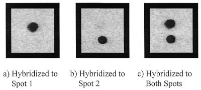
Images of DNA modified silicon (001) obtained using a Molecular Dynamics FluorImager. Two different oligonucleotides are attached to the surface in two spots ~2 mm across. Hybridization with the fluorescent complement of the upper spot shows the expected image (a). Denaturation and hybridization to the lower spot also shows the expected image (b). Denaturation and hybridization with both complements allows both spots to be visualized (c).
Control of amine surface density
Control of the density of surface attachment sites allows for control of the density of bound oligonucleotides. The surface density of the immobilized strands is an important parameter of the surface affecting DNA hybridization behavior (10) and amenability to enzymatic modification (54,55). Control of the surface density of amines was obtained in two different ways: (i) by using mixtures with varying mole ratios of protected amine and dodecene during the attachment step; and (ii) by controlling the degree of removal of the t-BOC protecting groups by varying the deprotection time.
Control of the density of surface-reactive amines was obtained using mixtures of t-BOC protected 10-aminodec-1-ene and dodecene. Dodecene is unreactive once attached to the silicon surface. Mixtures containing increasing fractions of t-BOC protected 10-aminodec-1-ene were applied to the surfaces and allowed to react under the UV lamp. The surfaces were then treated as before with TFA, SSMCC and DNA. Hybridization with the fluorescent complement and subsequent quantification of the fluorescence image was done on the surfaces. The plot in Figure 4 shows a linear relationship between the measured fluorescence signal and the fraction of 10-aminodec-1-ene. This experiment clearly demonstrates control of the density of surface amine groups available with a corresponding effect upon the surface density of bound oligonucleotides available for hybridization.
Figure 4.

Control of oligonucleotide density using binary mixtures of 10-aminodec-1-ene and dodecene. Mixtures containing different percentages of t-BOC protected 10-aminodec-1-ene and dodecene were attached to the surfaces using UV light. Deprotection and addition of SSMCC and thiol DNA followed by hybridization with the fluorescent complement allowed for quantification of the signal. A linear relationship is observed between the fluorescence signal obtained and the percent of amine attached to the surface (y = 0.0081x + 0.1664).
An alternate method of controlling the density of surface attachment sites involved controlling the exposure time of the t-BOC protected amine to deprotection conditions. Four surfaces were prepared with the t-BOC protected 10-aminodec-1-ene. The surfaces were then exposed to 25% TFA for 0, 2, 15 and 60 min, followed by modification with SSMCC and 5′ thiol DNA and hybridization with fluorescent complement. Figure 5 shows a plot of the fluorescence signals obtained from each of these surfaces. The amount of fluorescence recovered is dependent on the time of exposure to the deprotecting agent. Deprotection occurs rapidly up to 15 min when most of the t-BOC groups have been removed. Continuing the deprotection for 60 min yields a slight increase in the density of free amines. The plots of both Figures 4 and 5 show a non-zero intercept at 0% amine and 0 min of deprotection, respectively. A possible cause for this is that the SSMCC is non-specifically binding to the modified surfaces thereby allowing the subsequent attachment of a small amount of the thiol-modified oligonucleotides.
Figure 5.

Control of oligonucleotide density by exposing surfaces to deprotection conditions for differing lengths of time. Surfaces were deprotected, and then modified with SSMCC and thiol DNA. Hybridizing with the fluorescent complement followed by imaging on the FluorImager allowed for quantification of the signal. Exposure to TFA rapidly deprotects the t-BOC modified 10-aminodec-1-ene molecules to yield surface amines.
The density of fluorescent complement molecules hybridized to the surface bound oligonucleotides was determined using a previously published procedure (28,43). A DNA-modified surface was hybridized, then imaged to determine the total area of fluorescence. The surface was washed in 90°C water to denature and collect the fluorescent complements. The collected complements were loaded on one lane of a polyacrylamide gel while known amounts of fluorescent oligonucleotide standards were loaded in other lanes. After electrophoresis the gel was imaged and the amount of fluorescent oligonucleotide eluted from the surfaces was calculated by reference to a standard curve developed from known samples. Using this method the density of hybridized molecules was found to be ~2.3 × 10¹² molecules/cm2.
Stability
An important parameter of the DNA-modified silicon surfaces is their stability to the conditions employed for biochemical procedures such as DNA hybridization and enzymatic manipulation. Two experiments were performed to evaluate the stability of the DNA-modified surfaces. The stability of the bonds linking the DNA oligonucleotide covalently to the surface was evaluated by means of a fluorescent tag covalently attached to the oligonucleotide (S3); the surface was incubated in HB overnight at 27°C to ensure removal of non-specifically bound DNA and the surface fluorescence was measured repeatedly (Fig. 6). No significant decrease in the fluorescence intensity was observed, indicating that the DNA-surface linkage was stable under the conditions examined. In a second experiment, a surface modified with fluorescein labeled DNA (S3) in one area and non-labeled DNA (S1) in another was hybridized repeatedly with the fluorescent complement of S1. The measured fluorescence intensity of S3 was typically two-thirds that of S1 hybridized to its fluorescent complement. The normalized data show both signals decay at essentially identical rates (Fig. 7); the fluorescence intensity after 15 cycles is ~85% of the initial value, corresponding to a loss of ~1% per cycle. This stability is greater than that obtained in previously published results from our group using a non-covalent surface attachment chemistry, which showed a loss of 2% per cycle (43). These two experiments demonstrate that these DNA-modified silicon surfaces exhibit excellent stability under the storage and hybridization conditions employed.
Figure 6.

Stability of silicon surfaces covalently modified with fluorescently tagged oligonucleotides. DNA-modified silicon surfaces were prepared using 3′ thiol DNA with fluorescein on the 5′-end. The surfaces were stored overnight in HB then scanned with the FluorImager. Hourly scans recorded the surface fluorescence intensity. The observed fluorescence signal is stable throughout the experiment (y = –0.0041x + 0.9395).
Figure 7.

Stability of surfaces during 15 hybridization and denaturation cycles. The measured fluorescence signal from both the hybridized DNA and the fluorescein tagged DNA decreases by ~1% with each hybridization cycle. The surfaces were also imaged after each denaturation step to demonstrate the complete removal of the fluorescent complement. No fluorescence signal from the complement was observed after denaturation (hybridized DNA signal, y = –0.0097x + 1.0059; covalently attached DNA, y = –0.0076x + 0.6897).
The work reported here describes a method for covalently coupling oligonucleotides to silicon surfaces by functionalizing the surface with t-BOC protected 10-aminodec-1-ene and subsequently removing the acid-labile t-BOC group using 25% TFA in methylene chloride to produce the primary amine terminated surface. The progress of the surface modification reactions was monitored using contact angle and XPS measurements. Thiol-modified DNA was attached to the surface using the heterobifunctional crosslinker SSMCC. This method employs simpler and more direct coupling chemistry than previously described work (43) and provides control over the density of surface reactive sites. In addition, this surface chemistry was shown to provide excellent stability to hybridization and storage conditions.
REFERENCES
- 1.Rasmussen S.R., Larsen,M.R. and Rasmussen,S.E. (1991) Anal. Biochem., 198, 138–142. [DOI] [PubMed] [Google Scholar]
- 2.Yang M., McGovern,M.E. and Thompson,M. (1997) Anal. Chim. Acta, 346, 259–275. [Google Scholar]
- 3.Yang M., Kong,R.Y.C., Kazmi,M. and Leung,K.C. (1998) Chem. Lett., 3, 257–258. [Google Scholar]
- 4.Service R.F. (1998) Science, 282, 396–399. [DOI] [PubMed] [Google Scholar]
- 5.Kremski J.N., Wooters,J.L., Dougherty,J.P., Meyers,R.E., Collins,M. and Brown,E.L. (1987) Nucleic Acids Res., 15, 2891–2909. [DOI] [PMC free article] [PubMed] [Google Scholar]
- 6.Broude N.E., Sano,T., Smith,C.L. and Cantor,C.R. (1994) Proc. Natl Acad. Sci. USA, 91, 3072–3076. [DOI] [PMC free article] [PubMed] [Google Scholar]
- 7.Liu Q., Frutos,A.G., Thiel,A.J., Corn,R.M. and Smith,L.M. (1998) J. Comput. Biol., 5, 269–278. [DOI] [PubMed] [Google Scholar]
- 8.Henke L., Piunno,P.A.E., McClure,A.C. and Krull,U.J. (1997) Anal. Chim. Acta, 344, 201–213. [Google Scholar]
- 9.Maskos U. and Southern,E.M. (1992) Nucleic Acids Res., 20, 1679–1684. [DOI] [PMC free article] [PubMed] [Google Scholar]
- 10.Guo Z., Guilfoyle,R.A., Thiel,A.J., Wang,R. and Smith,L.M. (1994) Nucleic Acids Res., 22, 5456–5465. [DOI] [PMC free article] [PubMed] [Google Scholar]
- 11.Chrisey L.A., Lee,G. and O’Ferrall,C.E. (1996) Nucleic Acids Res., 24, 3031–3039. [DOI] [PMC free article] [PubMed] [Google Scholar]
- 12.Smith L.M., Corn,R.M., Condon,A.E., Lagally,M.G., Frutos,A.G., Liu,Q. and Thiel,A.J. (1998) J. Comput. Biol., 5, 255–267. [DOI] [PubMed] [Google Scholar]
- 13.Bamdad S. (1998) Biophys. J., 75, 1997–2003. [DOI] [PMC free article] [PubMed] [Google Scholar]
- 14.Parinov X., Barsky,V., Gennady,Y., Kirillov,E., Timofeev,E., Belgovkiy,A. and Mirzabekov,A. (1996) Nucleic Acids Res., 24, 2889–3004. [DOI] [PMC free article] [PubMed] [Google Scholar]
- 15.Livache T., Fouque,B., Roget,A., Marchand,J., Bidan,G., Teoule,R. and Mathis,G. (1998) Anal. Biochem., 255, 188–194. [DOI] [PubMed] [Google Scholar]
- 16.Southern E.M., Case-Green,S.C., Johnson,M., Mir,K.U., Wang,L. and Williams,J.C. (1994) Nucleic Acids Res., 22, 1368–1373. [DOI] [PMC free article] [PubMed] [Google Scholar]
- 17.Frutos A.G., Liu,Q., Thiel,A.J., Sanner,A.M.W., Condon,A.E., Smith,L.M. and Corn,R.M. (1997) Nucleic Acids Res., 25, 4748–4757. [DOI] [PMC free article] [PubMed] [Google Scholar]
- 18.Shchepinov M.S., Case-Green,S.C. and Southern,E.M. (1997) Nucleic Acids Res., 25, 1155–1161. [DOI] [PMC free article] [PubMed] [Google Scholar]
- 19.Wagner P., Nock,S., Spudich,J.A., Volkmuth,W.D., Chu,S., Cicero,R.L., Wade,C.P., Linford,M.R. and Chidsey,C.E.D. (1997) J. Struct. Biol., 119, 189–201. [DOI] [PubMed] [Google Scholar]
- 20.Arnold B.A., Hepler,R.W. and Keller,P.M. (1998) Biotechniques, 25, 106–110. [DOI] [PubMed] [Google Scholar]
- 21.Lund V., Schmid,R., Rickwood,D. and Hornes,E. (1988) Nucleic Acids Res., 16, 10860–10878. [DOI] [PMC free article] [PubMed] [Google Scholar]
- 22.Millan K.M., Spurmanis,A.J. and Mikkelsen,S.R. (1992) Electroanalysis, 4, 929–932. [Google Scholar]
- 23.Pang D., Zhang,M., Wang,Z., Qi,Y., Cheng,J. and Liu,Z. (1996) J. Electroanal. Chem., 403, 183–188. [Google Scholar]
- 24.Wang J., Fernandes,J.R. and Kubota,L.T. (1998) Anal. Chem., 70, 3699–3702. [DOI] [PubMed] [Google Scholar]
- 25.Millan K.M. and Mikkelsen,S.R. (1993) Anal. Chem., 65, 2317–2323. [DOI] [PubMed] [Google Scholar]
- 26.Zhao Y., Pang,D., Wang,Z., Cheng,J. and Qi,Y. (1997) J. Electroanal. Chem., 431, 203–209. [Google Scholar]
- 27.Hashimoto K., Ito,K. and Ishimori,Y. (1994) Anal. Chem., 66, 3830–3833. [DOI] [PubMed] [Google Scholar]
- 28.Frutos A.G., Smith,L.M. and Corn,R.M. (1998) J. Am. Chem. Soc., 120, 10277–10282. [Google Scholar]
- 29.Piunno P.A.E., Krull,U.J., Hudson,R.H.E., Damha,M.J. and Cohen,H. (1994) Anal. Chim. Acta, 288, 205–214. [Google Scholar]
- 30.Lamture J.B., Beattie,K.L., Burke,B.E., Eggers,M.D., Ehrlich,D.J., Fowler,R., Hollis,M.A., Kosicki,B.B., Reich,R.K., Smith,S.R., Varma,R.S. and Hogan,M.E. (1994) Nucleic Acids Res., 22, 2121–2125. [DOI] [PMC free article] [PubMed] [Google Scholar]
- 31.Souteyrand E., Cloarec,J.P., Martin,J.R., Wilson,C., Lawrence,I., Mikkelsen,S. and Lawrence,M.F. (1997) J. Phys. Chem. B, 101, 2980–2985. [Google Scholar]
- 32.Rogers Y., Jiang-Baucom,P., Huang,Z., Bogdanov,V., Anderson,S. and Boyce-Jacino,M.T. (1999) Anal. Biochem., 266, 23–30. [DOI] [PubMed] [Google Scholar]
- 33.Vo-Dinh T., Alarie,J.P., Isola,N., Landis,D., Wintenberg,A.L. and Ericson,M.N. (1999) Anal. Chem., 71, 358–363. [DOI] [PubMed] [Google Scholar]
- 34.Ulman A. (1996) Chem. Rev., 96, 1533–1554. [DOI] [PubMed] [Google Scholar]
- 35.Waddell T.G., Leyden,D.E. and DeBello,M.T. (1981) J. Am. Chem. Soc., 103, 5303–5307. [Google Scholar]
- 36.Linford M.R., Fenter,P., Eisenberger,P.M. and Chidsey,C.E.D. (1995) J. Am. Chem. Soc., 117, 3145–3155. [Google Scholar]
- 37.Buriak J.M. (1999) Chem. Commun., 12, 1051–1060. [Google Scholar]
- 38.Stewart M.P. and Buriak,J.M. (1998) Angew. Chem. Int. Ed., 37, 3257–3260. [DOI] [PubMed] [Google Scholar]
- 39.Sieval A.B., Demirel,A.L., Nissink,J.W.M., Linford,M.R., van der Mass,J.H., de Jeu,W.H., Zuilhof,H. and Sudholter,E.J.R. (1998) Langmuir, 14, 1759–1768. [Google Scholar]
- 40.Terry J., Linford,M.R., Wigren,C., Cao,R., Pianetta,P. and Chidsey,C.E.D. (1999) J. Appl. Phys., 85, 213–221. [Google Scholar]
- 41.Hamers R.J., Hovis,J.S., Seung,L., Liu,H. and Shan,J. (1997) J. Phys. Chem. B, 101, 1489–1492. [Google Scholar]
- 42.Effenberger F., Gotz,G., Bidlingmaier,B. and Wezstein,M. (1998) Angew. Chem. Int. Ed., 37, 2462–2464. [DOI] [PubMed] [Google Scholar]
- 43.Strother T., Cai,W., Zhao,X., Hamers,R.J. and Smith,L.M. (2000) J. Am. Chem. Soc., 122, 1205–1209. [Google Scholar]
- 44.Connolly B.A. and Rider,P. (1985) Nucleic Acids Res., 13, 4485–4501. [DOI] [PMC free article] [PubMed] [Google Scholar]
- 45. Glen Research Corporation (1996) User Guide to DNA Modification. Glen Research Corporation, Sterling, VA, pp. 22–23.
- 46.Pfister J.R. and Wymann,W.E. (1983) Synthesis, 1, 38–40. [Google Scholar]
- 47.Newman H. (1965) J. Org. Chem., 30, 1287–1288. [Google Scholar]
- 48.Tarbell D.S., Yamamoto,Y. and Pope,B.M. (1972) Proc. Natl Acad. Sci. USA, 69, 730–732. [DOI] [PMC free article] [PubMed] [Google Scholar]
- 49.Wong S.S. (1993) Chemistry of Protein Conjugation and Cross-Linking. CRC Press, Boca Raton, FL, pp. 7–48.
- 50.Hengge E. (1974) Properties and Preparations of Si-Si Linkages. Springer-Verlag, New York, NY, Vol. 51, pp. 10–47.
- 51.Hahn P.O. and Kerstan,M. (1988) SPIE Proc., 1009, 172. [Google Scholar]
- 52.Greene T.W. and Wuts,P.G.M. (1999) Protective Groups in Organic Synthesis. John Wiley & Sons, New York, NY, pp. 494–653.
- 53.Vogler E.A. (1993) In Berg,J.C. (ed.), Wettability. Marcel Dekker, New York, NY, Vol. 49, pp. 184–250.
- 54.Wang L., Liu,Q., Corn,R.M., Condon,A.E. and Smith,L.M. (2000) J. Am. Chem. Soc., 122, 7435–7440. [Google Scholar]
- 55.Bulyk M.L., Genalen,E., Lockhart,D.J. and Church,G.M. (1999) Nature Biotechnol., 17, 522–573. [DOI] [PubMed] [Google Scholar]