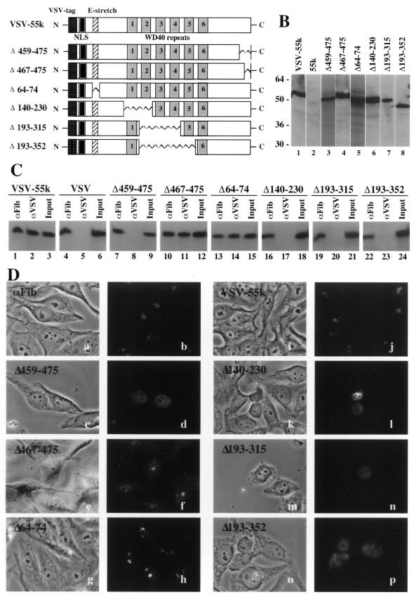
Figure 6.
U3-55k sequences required for association with U3 RNA and localization of U3-55k to the nucleolus. (A) The positions of the VSV tag, nuclear localization signals, glutamic acid-rich region and WD repeats in the VSV-tagged wild-type human U3-55k protein (VSV-55k) are represented with boxes. Regions of the protein deleted in each of the mutant constructs analyzed in this work are represented with zig-zag lines. (B) Expression levels of hU3-55k deletion mutants. Western blots using anti-VSV antibodies are shown for COS-1 cells expressing untagged hU3-55k (lane 2), VSV-tagged wild-type U3-55k (lane 1) and deletion constructs Δ459–475 (lane 3), Δ467–475 (lane 4), Δ64–74 (lane 5), Δ140–230 (lane 6), Δ193–315 (lane 7) and Δ193–352 (lane 8). Lanes 3 and 5 are taken from a different blot. (C) The effect of hU3-55k deletions on U3 snoRNP association. VSV-tagged human U3-55k wild-type and deletion constructs were expressed in COS-1 cells and complexes containing the tagged proteins (or endogenous fibrillarin) were immunoprecipitated using αVSV (or αFib) antibodies. Co-immunoprecipitated U3 RNA was detected by northern blotting. Co-immunoprecipitation of U3 with fibrillarin is shown for each experiment. The input lanes contain 10% of RNA isolated from extracts prepared for immunoprecipitation. (D) Cellular localization of hU3-55k wild-type and deletion mutants. VSV-tagged U3-55k wild-type and deletion constructs were expressed in HEp-2 cells and the localization of the tagged proteins (or endogenous fibrillarin) was assessed by immunofluorescence using αVSV (panels c–p) [or αFib (panels a and b)] antibodies. Phase contrast (panels a, c, e, g, i, k, m and o) and fluorescence (panels b, d, f, h, j, l, n and p) microscopy images are shown for each experiment. Fibrillarin is found primarily in the phase-dark nucleoli of HEp-2 cells (panels a and b). VSV-tagged wild-type U3-55k (panels i and j) and deletion constructs Δ467–475 (panels e and f) and Δ64–74 (panels g and h) were localized to nucleoli. Deletion mutants Δ459–475 (panels c and d), Δ140–230 (panels k and l), Δ193–315 (panels m and n) and Δ193–352 (panels o and p) did not localize to nucleoli but accumulated in the nucleoplasm.

