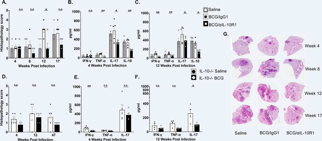
Figure 6. BCG/αIL-10R1 administration reduces immuno-pathology in lungs after M.tb infection.
WT mice were immunized with saline, BCG/αIL-10R1 or BCG/IgG1 (A-C). IL-10−/− CBA/J mice were vaccinated with saline or BCG (D-F). Mice were infected with M.tb 7- weeks post vaccination. WT and IL-10−/− mice were euthanized at predetermined time points post infection and the caudal lung lobe was quantified for pulmonary inflammation as percent of tissue involved. Pulmonary inflammation in (A) WT mice at 4, 8, 12 and 17 weeks post infection, (D) IL-10−/− mice at 4, 12 and 47 weeks post infection. Percent affected area was quantified by calculating the total area of the involved tissue over the total area of the lobe for each individual mouse and graded as 1, 2, 3, 4 and 5 which corresponded to <10%, <25%, 50%, <75%, >75% of affected tissue, respectively. (G) Representative images of hematoxylin and eosin stained lung sections of WT mice at 4, 8, 12 and 17 weeks post infection are shown to visualize tissue morphology. ELISA was performed on lung homogenates to measure the level of TNF-α, IL-17, IFN-γ and IL-10 in (B) WT mice at 4 weeks post infection, (C) WT mice at 12 weeks post infection, (E) IL-10 −/− mice at 4 weeks post infection and (F) IL-10 −/− mice at 12 weeks post infection (Data in Figure 6A–C represent the mean ± SE of one of two independent experiments with 3 to 5 mice in each group at each time point. Student’s t test was performed to determine statistical significance between BCG/αIL-10R1 and BCG/IgG1 (WT mice) or between saline and BCG (IL-10−/− mice), immunized experimental groups. *P<0.05; **P<0.01.

