
Figure 1.
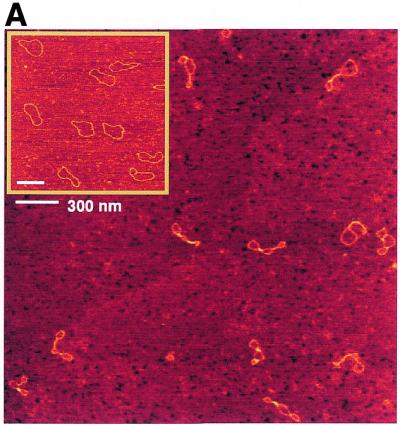
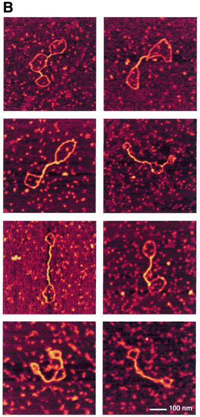
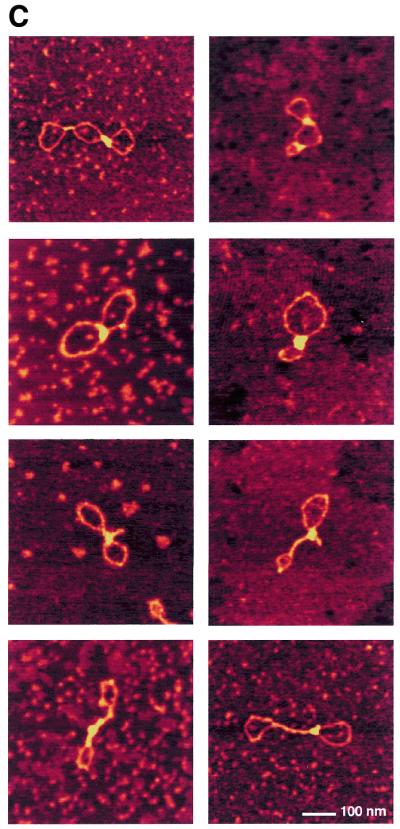
AFM images of circular nicked pUC19 molecules with and without H-NS. (A) DNA molecules after incubation with H-NS (1 dimer per 12 bp) on a 2.5 × 2.5 µm surface area and DNA molecules without H-NS on a 2 × 2 µm surface area (inset). (B) Close-up images of class I complexes. Complexes are laterally condensed and show a small reduction of DNA contour length (~3%). (C) Close-up images of class II complexes. Complexes show characteristic foci with a more dramatic level of condensation and a large reduction of DNA contour length (up to ~25%). All close-up images of condensed molecules show a 500 × 500 nm surface area. The colour scale ranges from 0.0 to 3.0 nm (from dark to bright).