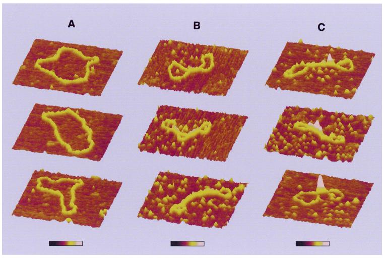
Figure 2.
Three-dimensional representation of circular nicked pUC19 molecules with and without H-NS. (A) DNA molecules without H-NS. (B) Class I complexes. (C) Higher order condensed class II complexes. Each series of images corresponds to characteristic structures found in that class. All images show a 400 × 400 nm surface area. The colour bar indicates sample height and corresponds to a 0.0–3.0 nm range (from dark to bright).

