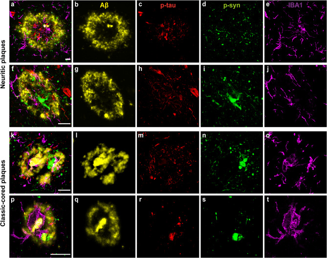
Fig. 5.
Representative confocal microscopy images of microglia in close proximity to AD-plaques in mixed DLB cases. Localization of pSer129-syn (green), Aβ (yellow), p-tau (red) and Iba1+ (purple) microglia in neuritic and classic cored plaques in the CA1, EntC and TC regions demonstrated with confocal microscopy. a-e Many large clustered and amoeboid Iba1-positive microglia with short, thick processes in the neuritic plaque in the CA1 within and in closer proximity to neuritic p-tau and p-syn accumulation. f-j Another example of rod-like and amoeboid Iba1-positive microglia in a neuritic plaque in the CA1. k-o Amoeboid and clustered microglia within and surrounding a classic-cored plaque in the EntC with neuritic p-tau and p-syn pathology. p-t Reactive microglial cell with large cell soma and thick processes within a classic-cored plaque with mainly Aβ pathology present in the TC. The scale bars in a, f, k, p are identical for all images in one row and represent 20 μm in all

