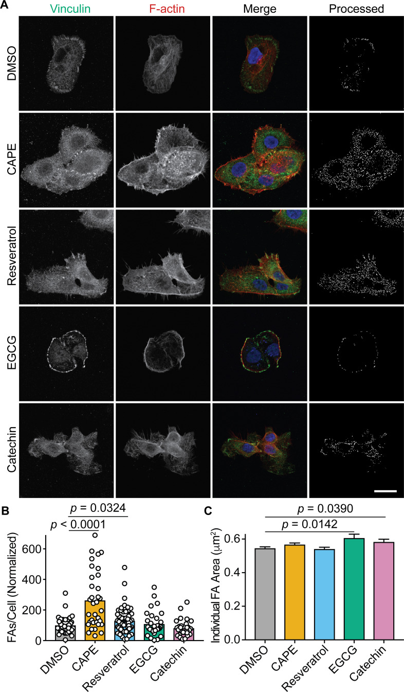
Fig 4.
CAPE and resveratrol increase focal adhesion numbers. (A) Confocal microscopy images of BECs that were treated for 3 h with carrier alone (DMSO), CAPE (25 µg/mL), resveratrol (22.9 µg/mL), EGCG (25 µg/mL), or catechin (25 µg/mL) and then fixed and stained for vinculin (green), F-actin (red), and nuclei (blue). Single-channel and merged images are indicated. The final panel in each row shows the cell images after processing to highlight focal adhesions for quantification. Scale bar, 10 µm. At least 30 cells from three independent experiments were processed to determine focal adhesion (B) numbers and (C) areas following the indicated treatments. Bars denote mean values (±SEM in C). P values were calculated relative to DMSO-treated controls by Student’s t tests.

