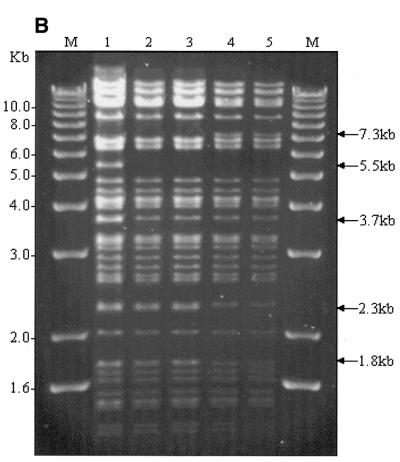
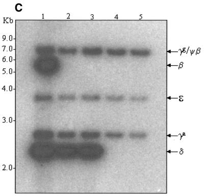
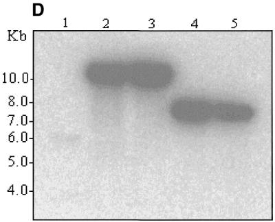
Figure 3.
(A) EcoRI restriction map in the region of the β- and δ-globin genes. (B) EcoRI digestion of unmodified and modified pEBAC 148β clones. Lane 1, unmodified pEBAC 148β; lanes 2 and 3, β globin gene replaced with the EGFP-Neo/Kan (two independent clones); lanes 4 and 5, δβ-globin genes replaced with the EGFP-Neo/Kan (two independent clones). (C) Southern blot analysis of the gel depicted in (B) and hybridised with a 32P-labelled LUG probe. (D) Southern blot analysis of the gel depicted in (B) and hybridised with a 32P-labelled EGFP-Neo/Kan probe.


