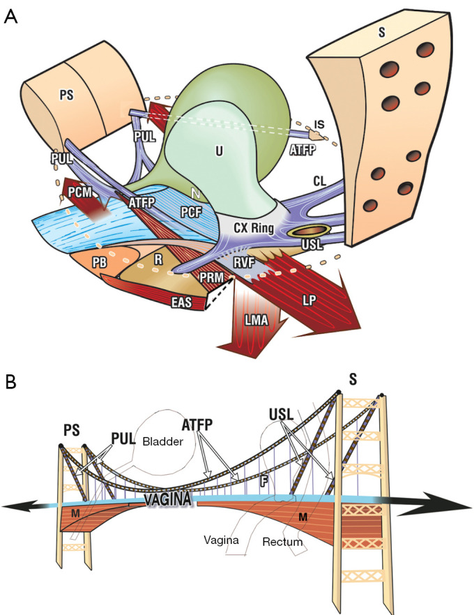
Figure 1.

The suspension bridge analogy for pelvic organ support. (A) The vagina is suspended by ligaments like a suspension bridge. Both walls of the vagina, PCF anteriorly, and RVF posteriorly, are suspended like the traffic part of a suspension bridge, by suspensory ligaments, PUL, CL, USL, ATFP laterally and PB inferiorly. The opposite muscle forces “M” (PCM, LP, and conjoint LMA), lie below the ligaments, and impart strength to the vaginal membrane by stretching it in opposite directions like a trampoline. (B) The suspension bridge analogy. Ligaments suspend the vagina from above and the pelvic muscles support it from below. The opposite muscle forces (arrows) stretch the vagina in opposite directions to tension it. Both images reused from Petros P. The female pelvic floor function, dysfunction and management according to the Integral Theory. 3rd ed. Heidelberg: Springer Berlin; 2010. With permission from Peter Petros; retains ownership of the copyright. PS, pubic symphysis; PUL, pubourethral ligament; PCM, pubococcygeus muscle; PB, perineal body; ATFP, arcus tendineus fascia pelvis; R, rectum; U, urethra; N, bladder base stretch receptors; PCF, pubocervical fascia; CX, cervix; RVF, rectovaginal fascia; PRM, puborectalis muscle; EAS, external anal sphincter; S, sacrum; IS, ischial spine; CL, cardinal ligament; USL, uterosacral ligament; LP, levator plate; LMA, longitudinal muscle of the anus; PS, pubic symphysis; M, muscle; F, fascia.
