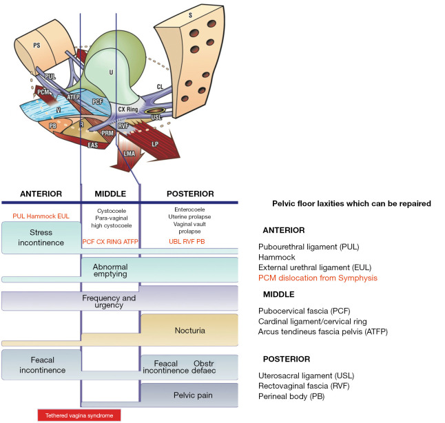
Figure 3.
Diagnostic algorithm. A “short-hand” diagnostic method where symptoms indicate which ligaments are causing which prolapse and which symptoms. The connective tissue structures fall naturally into three zones of causation. Symptoms, even if occurring “sometimes”, are ticked in each box where they occur. The ticked boxes also serve as a guide to surgery. For example, nocturia and pelvic pain are almost exclusively caused by the “USL” laxity; stress incontinence, by pubourethral laxity “PUL”. Reused from Petros P. The female pelvic floor function, dysfunction and management according to the Integral Theory. 3rd ed. Heidelberg: Springer Berlin; 2010. With permission from Peter Petros; retains ownership of the copyright. PS, pubic symphysis; PUL, pubourethral ligament; PCM, pubococcygeus muscle; V, vagina; PB, perineal body; ATFP, arcus tendineus fascia pelvis; R, rectum; U, urethra; PCF, pubocervical fascia; CX, cervix; RVF, rectovaginal fascia; PRM, puborectalis muscle; EAS, external anal sphincter; S, sacrum; IS, ischial spine; CL, cardinal ligament; USL, uterosacral ligament; LP, levator plate; LMA, longitudinal muscle of the anus; EUL, external urethral ligament.

