
Figure 3.
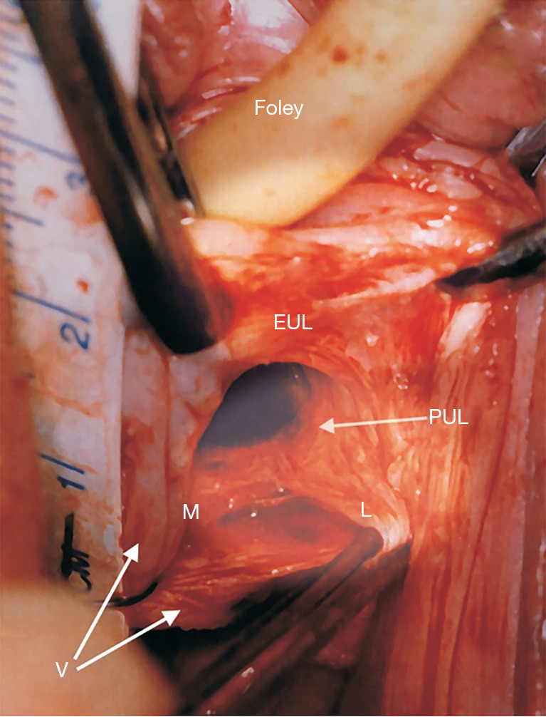
Live anatomy: surgical binding of loose PULs. Original live anatomical dissection of PUL from the two incision IVS operation (11). The tape measure overlies the urethra. The left paraurethral sulcus has been incised along its length and opened out laterally with forceps. “EUL” is the external urethral ligament which sits in front of the PS and is attached to the external urethral meatus. The PUL originates behind the PS, 1.5 cm from its lower border. Coming down from the PS, the PUL splits into two parts, medial (M) to insert into the side of the midurethra and L (lateral). “L” attaches laterally to pubococcygeus muscle (not seen) then down to attach to the vagina (V). Reused from Petros P. The female pelvic floor function, dysfunction and management according to the Integral Theory. 3rd ed. Heidelberg: Springer Berlin; 2010. With permission from Peter Petros; retains ownership of the copyright. EUL, external urethral ligament; PUL, pubourethral ligament; IVS, intravaginal slingplasty; PS, pubic symphysis.