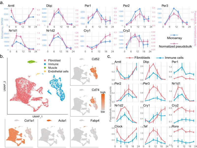
Fig. 4. The circadian clock is present in mouse dermal fibroblasts and immune cells.
a The normalized pseudobulk expression of the core clock genes generated from scRNAseq data (pink, n = 3 biologically independent samples per circadian time point) is consistent with their expression in the published microarray data (blue, n = 2 biologically independent samples per circadian time point, except that n = 3 at ZT2). Data are presented as mean values +/− SD. b Four major cell types, fibroblasts (red), immune cells (blue), muscle cells (green), and endothelial cells (yellow) were identified using canonical marker genes. Feature plots of the representative marker genes are shown (orange: high expression; grey: low expression); Col1a1 for fibroblasts, Acta1 for muscle cells, Fabp4 for endothelial cells, Cd52 and Cd74 for immune cells. c At the pseudobulk-level, expression pattern of the core clock genes is similar in fibroblasts (red) and immune cells (blue), while the amplitudes of the oscillations are dampened in immune cells for most of the core clock genes. n = 3 biologically independent samples per circadian time point. Data are presented as mean values +/− SD. Source data are provided as a Source Data file.

