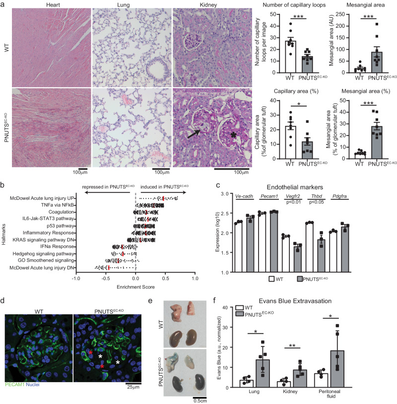
Fig. 3. Induction of endothelial-specific PNUTS KO in mice provokes vascular leakage and multiorgan failure.
a Histopathological study of different tissues in PNUTSEC-KO mice compared to WT mice: left, heart samples (haematoxylin-eosin) presenting edema; middle, lung samples (haematoxylin-eosin) presenting thrombi; right, kidney samples (PAS staining) showing glomerulosclerosis (asterisk) and capillary dilatation (black arrow) in renal glomeruli. Quantifications of the kidney sections show the absolute and relative contribution of capillaries and mesangial tissue in glomeruli (n = 8 per group). b RNA-seq was performed with lung tissue of WT and PNUTSEC-KO mice and was analysed for differentially regulated pathways using Gene Set Enrichment Analysis. Enrichment scores of the indicated pathways are plotted on the x axis (n = 3). c The expression levels of the endothelial markers Ve-cadherin, Pecam1, Vegfr2, Thbd and Pdgfra in WT and PNUTSEC-KO lung samples were confirmed by RT-qPCR and normalized to Rplp0 mRNA (n = 3). d The presence of ECs in the glomerular capillary network was investigated by PECAM1 immunostaining in kidneys of WT and PNUTSEC-KO mice. Red and white asterisks indicate potential mesangial proliferation and glomerulosclerotic areas, respectively. e, f An Evans Blue (EB) extravasation assay was performed to measure the vascular extravasation in different organs. e Representative lung and kidney images 1 h after intravenous administration of EB at 25 mg/kg. f Colorimetric measurement of extravasated EB into lung, kidney and peritoneal fluid (n = 4–5 mice per group). *p < 0.05, **p < 0.01, ***p < 0.001. Error bars depict the standard error of the mean (SEM).

