Figure 1.
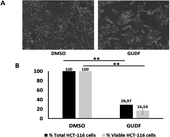
GUDF Induces Proliferative Arrest and Cell Death in HCT-116 Colon Cancer Cells. (A) HCT-116 colon cancer cells were treated by DMSO and GUDF (50 µg/ml) for 72 hr. Routine microscopy was performed at 100X to observe the cell number and cellular morphology. (B) HCT-116 colon cancer cells were treated by DMSO and GUDF (50 µg/ml) for 72 hr. Percentage total cell and viable HCT-116 colon cancer cells estimated by Trypan blue dye exclusion assay. Trypan blue dye exclusion assay was performed to estimate viable and dead cells. Data are represented as mean ± SD. Each experiment was conducted independently three times. The bar graph without an asterisk denotes that there is no significant difference compared to the DMSO control. * Significantly different from DMSO control at the P-value < 0.05. ** Significantly different from DMSO control at P-value < 0.01. *** Significantly different from DMSO control at P-value < 0.001

