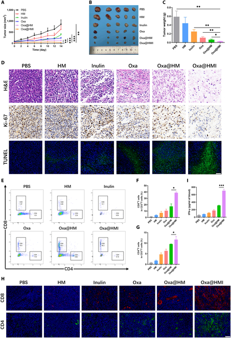
Fig. 10.

In vivo antitumor activity and the tumor immune microenvironment modulation by Oxa@HMI Gel on subcutaneous colorectal tumor-bearing Balb/c mice. (A) The tumor growth curves of subcutaneous colorectal tumor-bearing mice received various treatments (n = 5). Representative images (B) and weight (C) of tumors excised from the sacrificed mice after medication process (n = 5). (D) Histological analysis by H&E, Ki-67, and TUNEL staining of tumor sections after various treatments. Scale bar: 20 μm. Representative flow cytometric images (E) and corresponding quantitative analysis of CD8+ T cells (F) and CD4+ T cells (G) infiltrated in the tumor tissues form mice after different treatments (n = 3). (H) Immunofluorescent staining images of CD8+ T cells and CD4+ T cells in tumor tissues from mice after various treatments. Scale bar: 20 μm. (I) The secretion levels of serum IFN-γ determined by enzyme-linked immunosorbent assay (n = 3). The data are presented as mean ± SD. *P < 0.05, **P < 0.01, ***P < 0.001.
