. 2024 Apr 2;15(18):6752–6762. doi: 10.1039/d4sc01214c

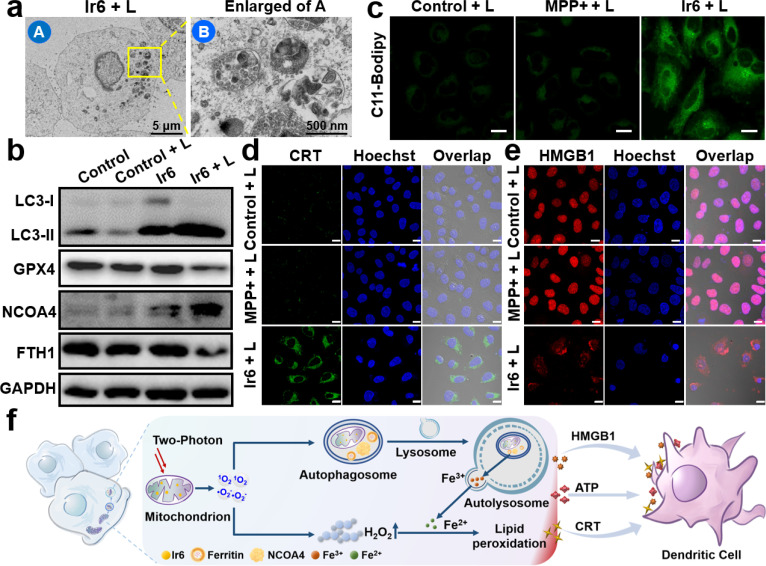
Fig. 3. Cell death mechanism of Ir6 in A375 cells. (a) Representative transmission electron microscopy image of the autophagosomes of A375 cells upon treatment with Ir6 upon irradiation (L). (b) Western blot analysis of the expression level of LC3-I, LC3-II, GPX4, NCOA4, and FTH1 of A375 cells upon treatment with Ir6 in the dark or irradiation (L). (c) CLSM of A375 cells incubated with the lipid peroxide-specific probe C11-Bodipy and treated with MPP+ or Ir6 upon irradiation (L). Scale bar = 20 μm. Immunofluorescence CLSM of A375 cells incubated with (d) CRT-specific antibody and (e) HMGB1 protein-specific antibody and treated with MPP+ or Ir6 upon irradiation (L). Scale bar = 20 μm. (f) Proposed cell death mechanism of ferritinophagy and immunogenic cell death by Ir6 upon irradiation. Concentration: Ir6 (10 μM), MPP+ (100 μM). L = two-photon laser irradiation at 720 nm, 40 mW, 120 s.

Fig. 3