. 2024 Apr 2;15(18):6752–6762. doi: 10.1039/d4sc01214c

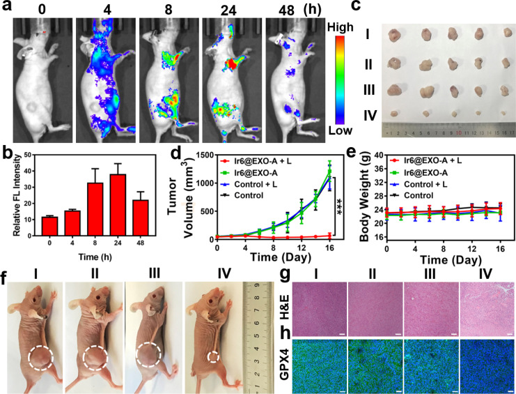
Fig. 4. (a) Time-dependent luminescence photographs of an A375 tumor-bearing mouse model after injection of Ir6@EXO-A into the tail vein in an animal imaging system. (b) Determination of the time-dependent fluorescence in the tumor, n = 3. (c) Photographs of the tumor of an A375 tumor-bearing mouse model 16 days after treatment. (d) Tumor growth inhibition curves of an A375 tumor-bearing mouse model during the treatment. (e) Changes in the body weight of an A375 tumor-bearing mouse model during the treatment. (f) Representative photos of the xenograft model of A375 tumors after different treatments. (g) Hematoxylin and eosin stain (H&E) of the tumorous tissue of an A375 tumor-bearing mouse model 16 days after treatment. (h) Glutathione peroxidase 4 stain (GPX4) of the tumorous tissue of an A375 tumor-bearing mouse model 16 days after treatment. I: control, II: control + L, III: Ir6@EXO-A, IV: Ir6@EXO-A + L. L = two-photon laser irradiation at 720 nm, 50 mW, 300 s. Scale bar = 50 μm. Error bars = standard deviation, n = 5. ***p < 0.001.

Fig. 4