
Figure 4.
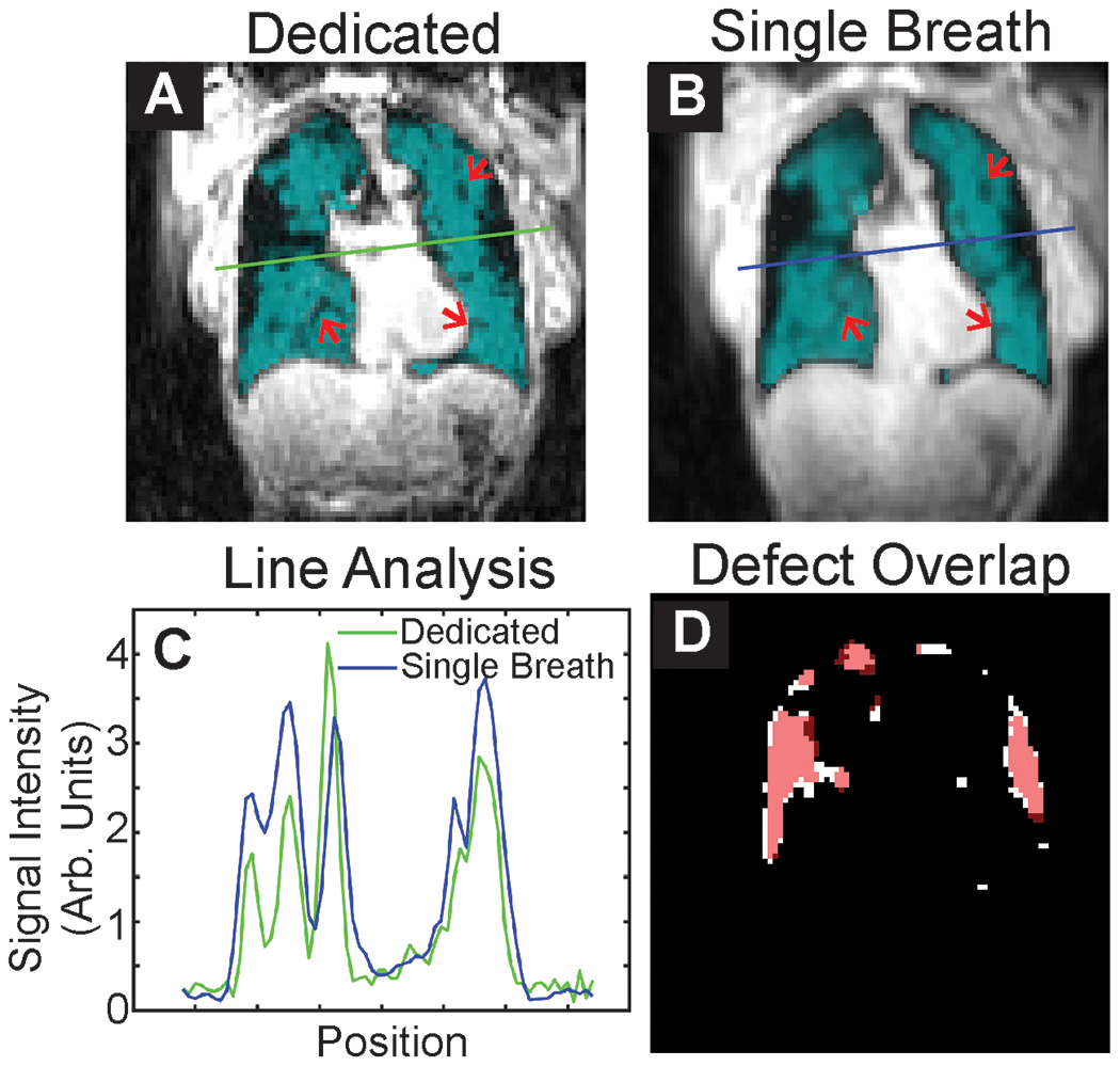
Representative slices from A Dedicated and B Single-breath ventilation imaging. Subtle ventilation defects visible in both images are highlighted by arrows. C Line analysis shows slightly greater blurring of images for single-breath images, which is expected given the lower resolution of these images (4.2 vs. 4 mm nominal resolution). D Overlap of defect maps, with dedicated shown in white and single-breath shown in red. There is considerable overlap of ventilation defects.