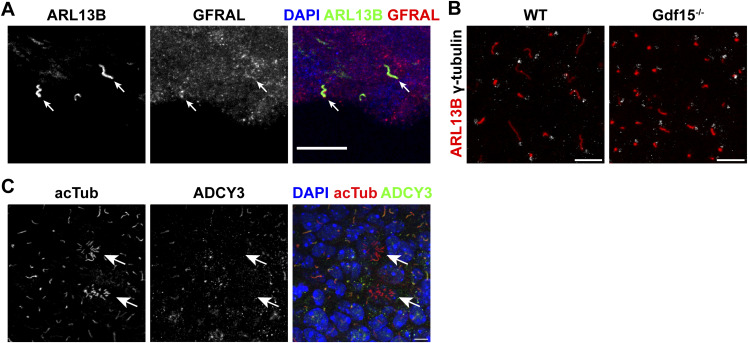
Figure S1. Characterization of primary cilia in the GE/V-SVZ.
(A) Immunofluorescent micrographs of the lateral SVZ in coronal sections of adult WT mice, labelled for cilia marker ARL13B (green), and GFRAL (red). DAPI was used for nuclear counterstain. Scale bar = 20 μm. (B) Immunofluorescent micrographs of whole mounts of the E18 GE labelled for ARL13B and γ-tubulin show no difference in γ-tubulin localization in relation to the cilium. Scale bars = 10 μm. (C) Immunofluorescent micrographs of whole mounts of the E18 GE labelled for acTub and ADCY3 showing two emerging ependymal cells (white arrows), demonstrating a clear difference between primary and emerging motile cilia. DAPI was used for nuclear counterstain. Scale bar = 5 μm.

