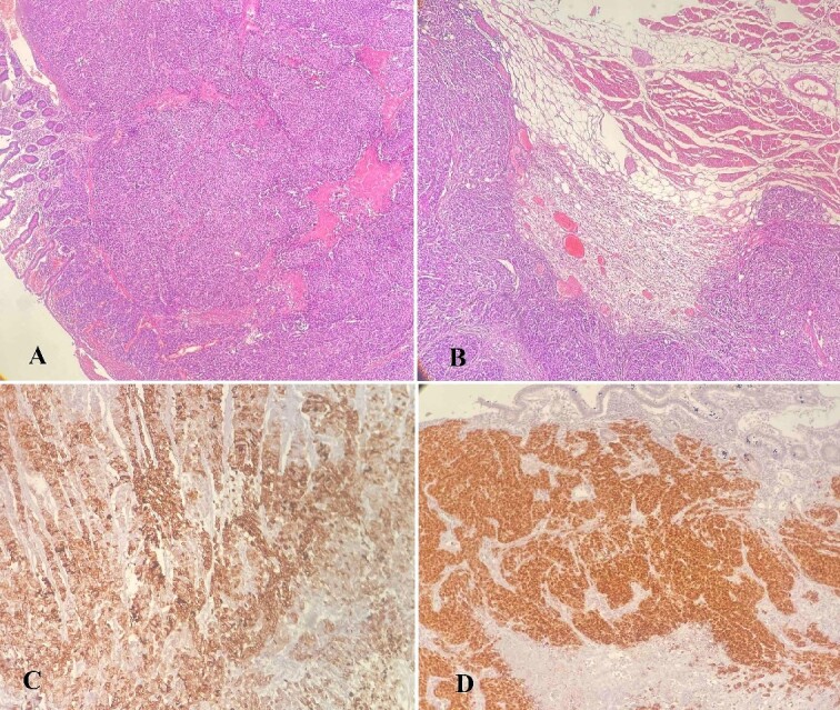
Figure 6.

(A–D) Histology analysis; (A) tumor cells infiltrating the wall of the small intestine with mucosal ulceration (hematoxylin–eosin stain, ×40); (B) neoplastic cells infiltrating skeletal muscle of the abdominal wall (hematoxylin–eosin stain, ×40); (C) positive staining for CK7, ×100; (D) positive staining for PAX8, ×100.
