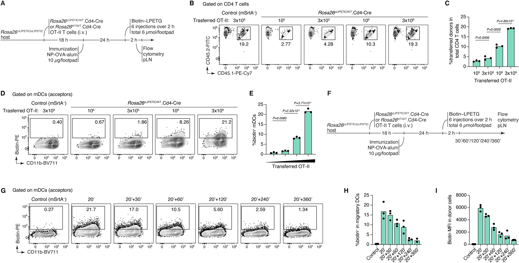
Extended Data Figure 3 |. uLIPSTIC labeling of T cell–DC interactions in adaptive transfer models.

(A-E) mSrtA+ donor cell numbers determine the degree of uLIPSTIC labeling. (A) Experimental layout for panels (B-E). Increasing numbers (105, 3 × 10⁵, 106, 3 × 10⁶) of Rosa26uLIPSTIC/+.CD4-Cre OT-II CD4+ T cells were adoptively transferred into recipient Rosa26uLIPSTIC/ uLIPSTIC mice, followed by OVA/alum immunization 18 h post-transfer and LIPSTIC substrate injection one day later. The number of transferred cells (CD45.1/2) determined the proportion of donor cells in the CD4+ T cell compartment (B-C) and the corresponding percentage of labeled interacting cells in the mDC compartment (D-E). (F-I) Persistence of label on acceptor cells with time. (F) Experimental layout for panels (G-I). (G) uLIPSTIC labeling of mDCs after incremental delays between substrate injection and tissue harvest. Quantified in (H,I). Data for all plots are for three mice per condition from one experiment. P-values were calculated using two-tailed Student’s tests.
