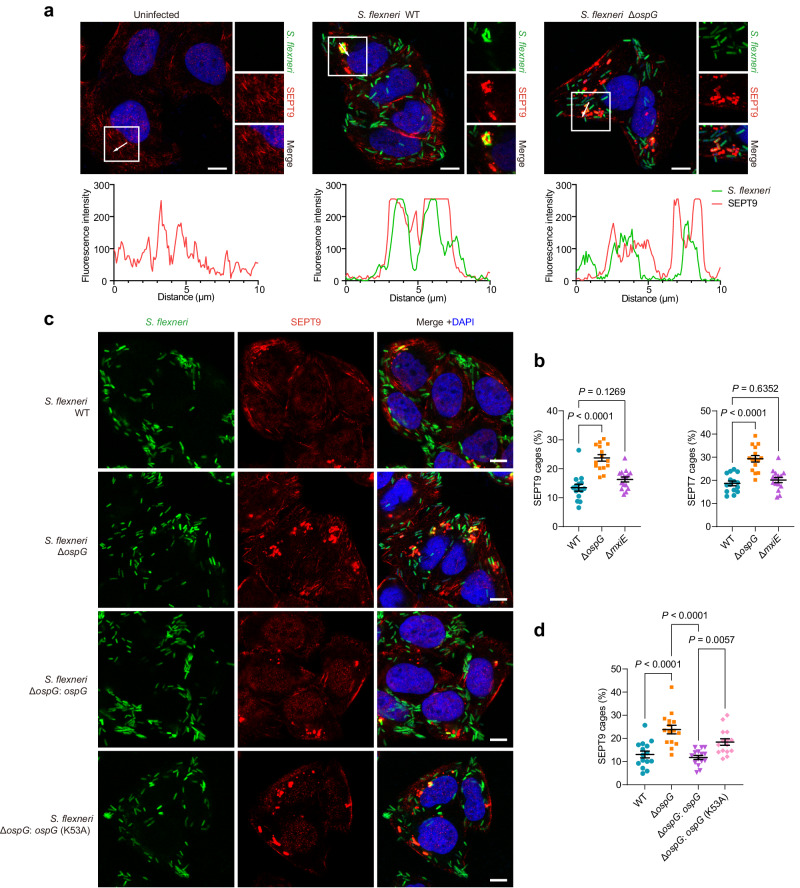
Fig. 6. OspG-promoted ubiquitination of septins prevents the assembly of septin-cages around cytosolic S. flexneri in infected host cells.
a Representative immunofluorescence images of uninfected or S. flexneri-infected cells. HeLa cells were infected with indicated GFP-labeled S. flexneri strains for 2 h, fixed and stained with an anti-SEPT9 antibody. Nuclei were stained with DAPI. Fluorescence intensity was plotted along the arrows. Scale bars, 10 μm. b HeLa cells were infected with S. flexneri at an MOI of 10 for 2 h, fixed and stained with anti-SEPT9 or anti-SEPT7 antibodies for quantitative microscopy. Data represent the mean % ± SEM of S. flexneri inside septin-cages from n = 3 biologically independent experiments and a total of 150 HeLa cells in which the proportion of septin-cages was counted. One-way ANOVA followed by Dunnett’s multiple comparison test was performed. c Representative immunofluorescence images of HeLa cells infected with indicated GFP-labeled S. flexneri (MOI = 10) strains for 2 h, fixed and stained with an anti-SEPT9 antibody. Nuclei were stained with DAPI. Scale bars, 10 μm. d HeLa cells were infected with S. flexneri at an MOI of 10 for 2 h, fixed and stained with an anti-SEPT9 antibody for quantitative microscopy. Data represent the mean % ± SEM of S. flexneri inside septin-cages from n = 3 biologically independent experiments and a total of 150 HeLa cells in which the proportion of septin-cages was counted. One-way ANOVA followed by Dunnett’s multiple comparison test was performed.

