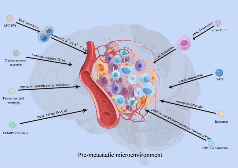
Figure 2.
Functions of EVs in developing pre-metastatic microenvironment. EVs derived from antigen presenting cells (APCs) can also activate CD8+and CD4+ T cells. Exosomes carrying PD-L1 promoted tumor growth and reduced the number of T cells in the spleen and lymph nodes in mouse experiments. Integrins (ITGs) fuse with target cells in a tissue-specific manner to direct organ-specific colonization. Tumour-secreted CEMIP+ exosomes uptaked by brain endothelial and microglial cells, upregulating the pro-inflammatory cytokines encoded by Ptgs2, Tnf and Ccl/Cxcl, promote brain vascular remodelling and metastasis. Human brain microvascular endothelial cells (HBMECs)-derived exosomes induce the elevated S100A16 in SCLC brain metastasis, and the S100A16-mediated protective effect is related to the up-regulation of prohibitin (PHB)-1.

