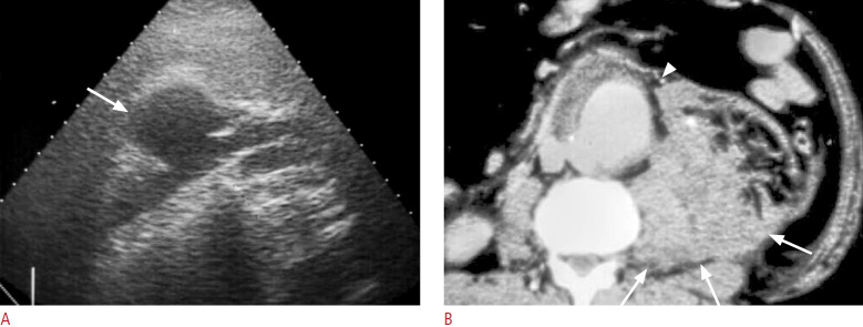
Fig. 13. Mycotic aneurysm of the infrarenal aorta complicated with rupture.
A. Left coronal sonography of the retroperitoneum shows a round hypoechoic mass due to abdominal aortic aneurysm with wall interruption (arrow). The aneurysm is associated with surrounding soft tissue or hematoma. B. Axial view of contrast-enhanced computed tomography shows rupture of the mycotic aortic aneurysm with wall interruption (arrowhead) and retroperitoneal hematoma (arrows).

