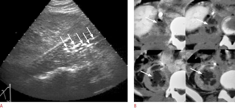
Fig. 18. A case of retroperitoneal abscess in the psoas muscle.
A. Right coronal ultrasonography shows multiple echogenic foci (arrows) in the right psoas muscle due to gas. B. Sequential axial view of contrast-enhanced computed tomography reveals a gas-containing abscess in the right psoas muscle (arrows).

