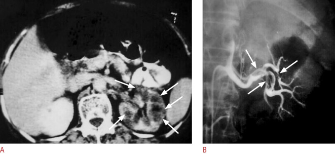
Fig. 21. A case of acute left flank pain with hematuria due to emboli from the left cardiac atrium.
A. Contrast-enhanced computed tomography shows hypodense areas (arrows) in the left renal parenchyma which is consistent with renal infarct. The findings were not detected by ultrasound (not shown). B. Transcatheter left renal angiography displays filling defects (arrows) in the left main and segmental renal arteries.

