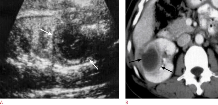
Fig. 5. A case of renal abscess.
A. Ultrasonography of the right kidney shows a well-defined hypoechoic mass (white arrows) due to renal abscess. B. Contrast-enhanced computed tomography of the right kidney shows a well-defined hypodense mass with an enhanced wall (black arrows) due to renal abscess.

