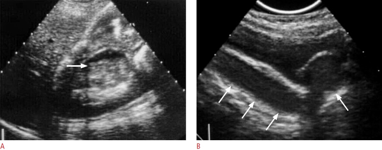
Fig. 6. A case of pyonephrosis and pyoureter.
A. Ultrasonography of the right kidney shows dilated right upper collecting system with fluid-fluid layering (arrow) suggesting pyocalyx. B. Sagittal ultrasonography of the pelvis in another patient shows ureterocele and pyouretrer with internal echoes due to suppurative echogenic debris (arrows) in the obstructed urotract suggesting pyonephrosis and pyoureter.

