
Fig. 8. A case of type II emphysematous pyelonephritis.
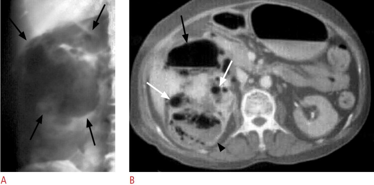
A. Radiography of the abdomen shows loculated and bubbly gas (arrows) in the right renal area. B. Computed tomography with a modified lung window level shows gas in collecting system (white arrows) as well as gas and fluid content in the right kidney (black arrowhead) and perirenal space (black arrow).