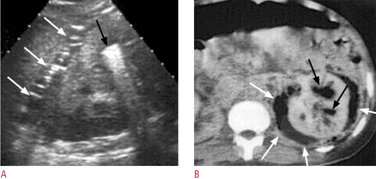
Fig. 9. A case of type II emphysematous pyelonephritis.

A. Ultrasonography shows echogenic foci in the left renal calyx (black arrow) and perirenal space (white arrows) due to gas content. B. Post-enhanced computed tomography shows gas in the collecting systems (black arrows) and perirenal space (white arrows) with subtle fluid content.
