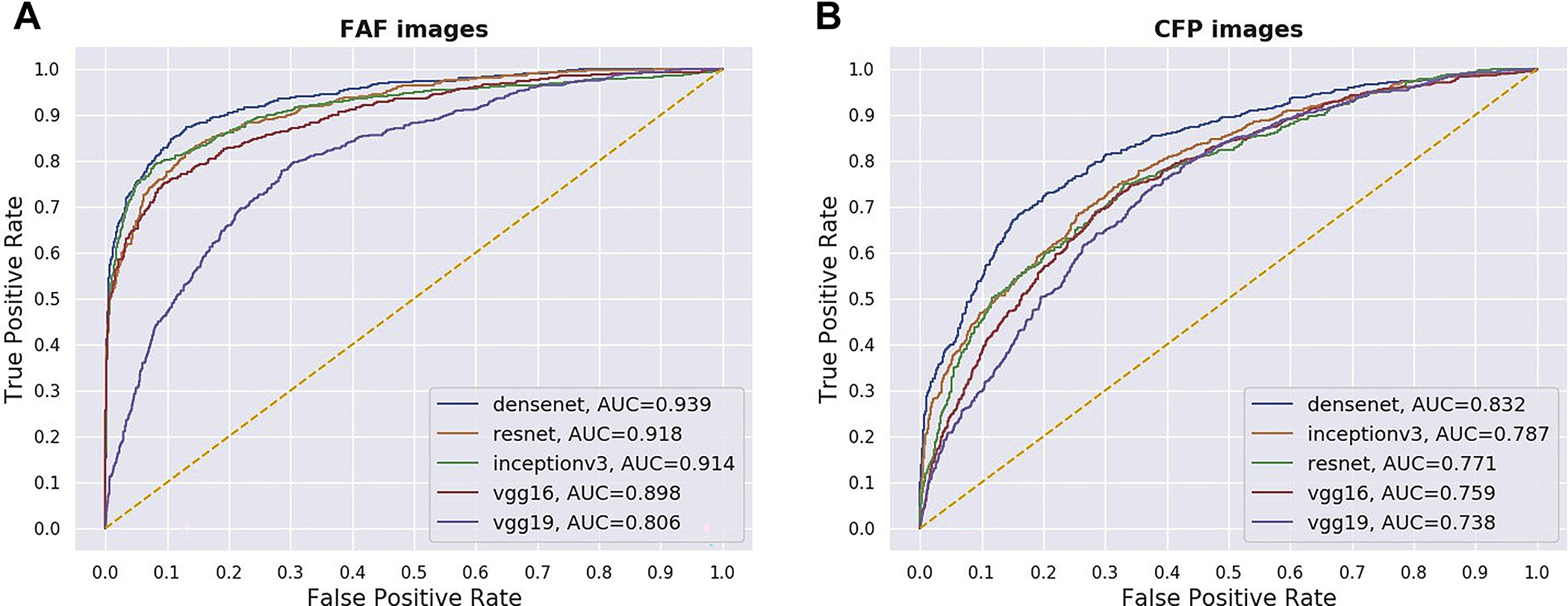
Figure 2.

Receiver operating characteristic curves of 5 different deep learning convolutional neural networks for the detection of reticular pseudodrusen from (A) fundus autofluorescence (FAF) images and (B) their corresponding color fundus photography (CFP) images, using the full test set. AUC = area under the receiver operating characteristic curve.
