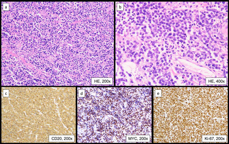
Fig. 3.
Histopathological examinations of a peritoneal biopsy specimen: (a) hematoxylin and eosin staining (H&E) ×200, (b) H&E ×400, (c) CD20 ×200, (d) MYC ×200, (e) Ki-67 labeling index ×200. Diffuse proliferation of medium-sized cells with an increase in the nuclear: cytoplasmic ratio is observed. These cells are relatively uniform in size and have hyperchromatic nuclei without conspicuous nucleoli. Coarse nuclear chromatin and marked nuclear pleomorphism, which are often observed in typical DLBCL, are not prominent (a, b). Tumor cells are positive for CD20 and MYC (c, d). The Ki-67 labeling index is approximately 95% (e).

