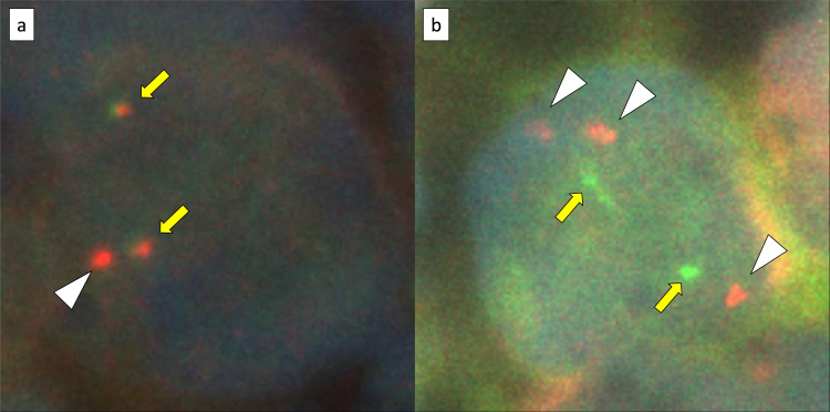
Fig. 4.
MYC gene status analysis by fluorescence in situ hybridization using formalin-fixed paraffin-embedded tissues: (a) the Vysis LSI-MYC dual color break-apart rearrangement probe (Abbott Laboratories), which contains LSI-MYC SpectrumOrange and LSI-MYC SpectrumGreen probes. The LSI-MYC SpectrumOrange probe begins 119 kb centromeric to the 5’ of the MYC gene and extends 260 kb towards the centromere. The LSI-MYC SpectrumGreen probe starts approximately 1.5 Mb telomeric to the 3’ of MYC gene and extends towards the telomere for about 400 kb. (b) the Vysis LSI IGH/MYC dual fusion probe (Abbott Laboratories), which contains an approximately 821 kb of the SpectrumOrange probe and an approximately 1.6 Mb of the SpectrumGreen probe, which cover the MYC and IGH regions, respectively. The MYC break-apart probe shows an extra red signal (white arrow head) besides two sets of red and green fusion signals (yellow arrow) in 84.0% of the cells (a). The IGH-MYC dual-fusion probe shows no fusion signal, however, three isolated red signals (white arrow head) and two green signals (yellow arrow) are observed in most analyzed cells (b).

