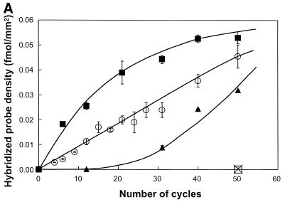
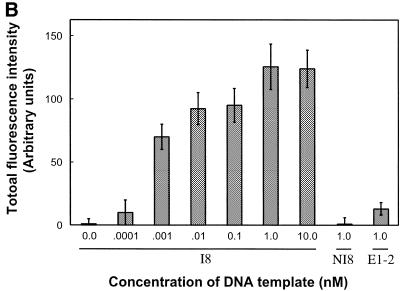
Figure 4.
Effect of the concentration of DNA template in solution. A 50 µM primer solution (5′-SH up1 and 5′-SH up2) was applied on glass slides derivatised with ATS and functionalised with the bi-functional cross-linker s-MBS. (A) Glass slides were submitted to solid phase PCR with increasing concentrations of template I8: 0.1 nM (triangle), 1 nM (circle), 10 nM (square). The loading density of DNA molecules (fmol/mm2) amplified on glass surface was determined by hybridisation with radioactively-labelled probe. The negative control, 1 nM of NI8 template (open square), has the same sequence as I8 but does not contain up1 or up2 primer sequences. To check the specificity of the hybridisation assay a non-specific radioactive probe was used (cross). (B) Fifty PCR cycles were run with I8 template (0, 0.0001, 0.001, 0.01, 0.1, 1 and 10 nM) or non-specific templates (1 nM of NI8 or E1-2). Glass slides were submitted to hybridisation using specific I8 digoxygenin-labelled long probe and the fluorescence signal was measured using the epi-fluorescence microscope.


