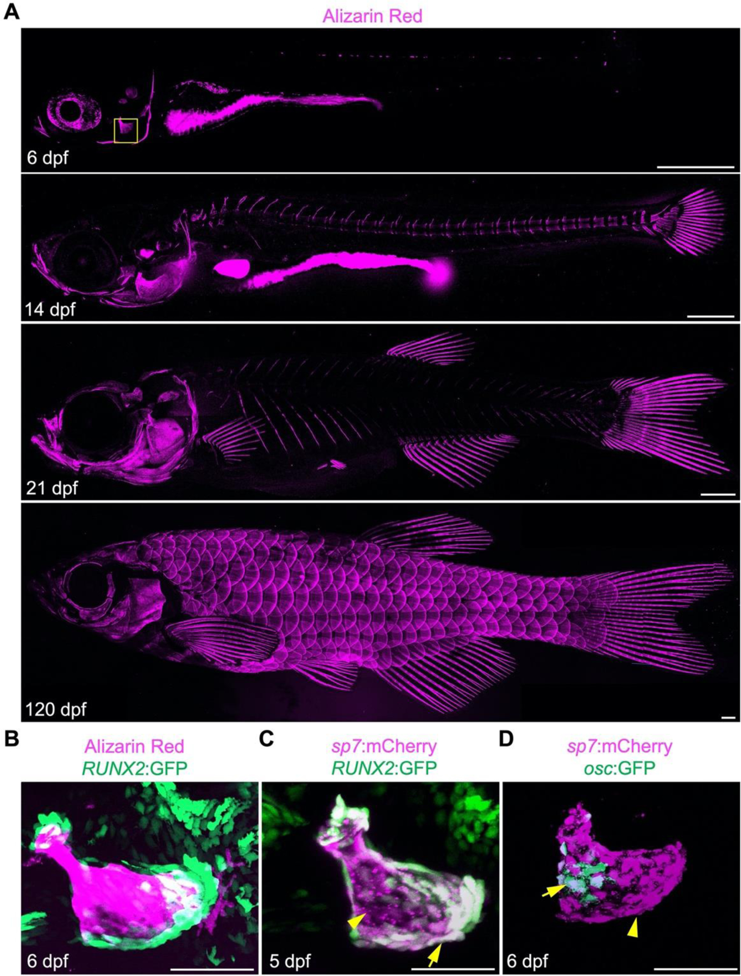
Figure 2.

Bone development in zebrafish. (A) Developmental series of alizarin red stained zebrafish from larval, juvenile and adult stages. Mineralized bones are detected earliest in the craniofacial skeleton and later appear within the axial and fin skeleton. Scales emerge later in development. Scale bars, 500 μm. (B-D) Osteogenesis in the larval opercula (boxed in A at 6 dpf). (B) RUNX2:GFP preosteoblasts are enriched at the tips of the mineralized larval opercular. (C) sp7:mCherry high cells are enriched along the surface of the opercular, while sp7:mCherry; RUNX2:GFP pre-osteoblast are abundant at the bone tips. (D) osc:GFP+ osteoblasts are restricted to the most mature osteoblasts away from the edges of the opercule. Scale bars for B-D, 50 μm.
