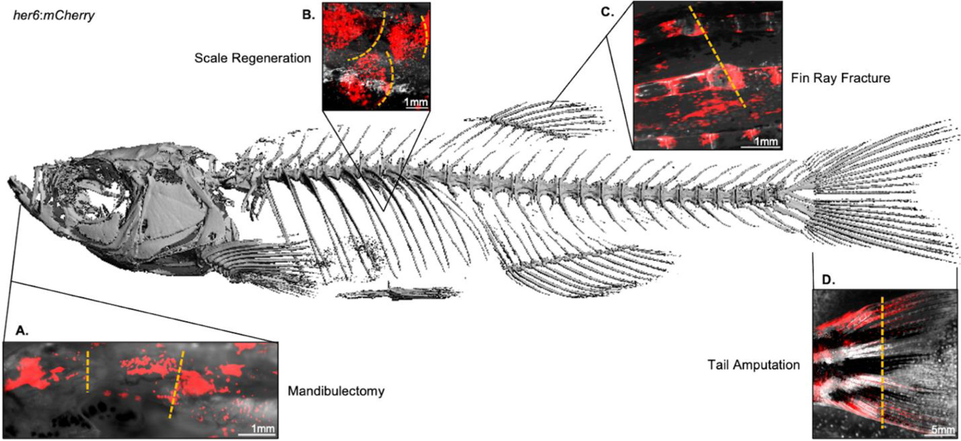
Figure 4.

Diverse regenerative processes activate notch signaling in the zebrafish skeleton. Micro-computed tomography reconstruction of the adult zebrafish skeleton, obtained in a Scanco μCT40 at 12μm^3 resolution with a lower threshold of 250 mgHA/ccm. Microscopy of zebrafish expressing the Notch signaling reporter her6:mCherry demonstrates ability to perform longitudinal intravital imaging of fluorescent transgenes under anesthesia using a stereomicroscopy. Representative images are shown 10 days following each of the following model injuries: (A) unilateral mandibulectomy; (B) scale plucking; (C) dorsal fin ray fracture; and (D) partial tail amputation, which were performed in different animals. Yellow dashed lines in A-D represent approximate injury margins.
