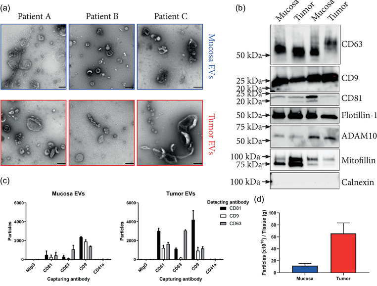
FIGURE 2.

Analysis of tissue Isolates. (a) Electron micrographs showing the vesicle isolates of three patients from both mucosa and tumour tissue. These three patient samples were not included in the proteomics analysis. Scale bars = 200 nm. (b) Tumour and mucosa isolates of two patients were probed for CD9, CD63, CD81, Flotillin 1, ADAM10, Mitofilin and Calnexin by western blot. (c) ExoView analysis of EVs captured by immobilised antibodies against CD9, CD63, CD81 and CD41a and subsequently probed with fluorescent antibodies against CD9, CD63 and CD81 showing the presence of EVs that were double positive for these markers. CD9, CD63 and CD81 are EV markers, while CD41 served as a platelet marker indicating the non‐detectable contamination of blood that could have occurred during surgery. MIgG serves as an isotype control (n = 1, error bars = SD for the technical replicates on the chip). (d) Particle measurement of three paired mucosa and tumour isolates showed an increase of particles in relation to tissue weight for tumour tissue isolates with 11.9 × 10¹⁰ ± 3 × 10¹⁰ and 65.9 × 10¹⁰ ± 14 × 1010 particles/gram tissue for mucosa and tumour, respectively (n = 3, error bars = SD).
