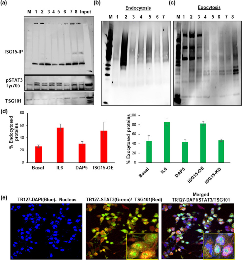
FIGURE 3.

ISGylation of STAT3 and its interaction with ISG15 modulate the vesicle secretion. (a) Pull down of ISG15 by immunoprecipitation in different POCC cells cultured from different patient ascites (Lane 2‐8) along with Input and Marker lane (M) shows the ISGylation of STAT3 and TSG101,a MVB protein, when compared with immortalized HGSOC cancer cells (OVCAR4‐ Lane1) at 24 h after culture. (b) Cell surface biotinylation assay: Endocytosis in TR127 cells at 24 h shows the influence of STAT3 and ISG15 OE and inhibition in protein endocytosis in Basal (Lane 4), IL6 treated cells for STAT3 activation (Lane 5), ISG15 inhibition by DAP5 treatment (Lane 6), and ISG‐OE cells (Lane 7) as compared to their controls‐untreated (Lane 1), biotinylating control (Lane 2), strip control (Lane 3) and marker lane (M). (c) Exocytosis measured in TR127 cells at 24 h in different treatment conditions in Basal (Lane 4), IL6 treatment (Lane 5), ISG15 inhibition by DAP5 treatment (Lane 6), ISG‐OE cells (Lane 7) and ISG15 KD cells (Lane 8) as compared to their controls‐untreated (Lane 1), biotinylating control (Lane 2), strip control (Lane 3) and marker lane (M). (d) Graph showing the % of endo‐ and exocytosed proteins in different treatment conditions described above. (e) Confocal microscopy showing co‐localization of STAT3 and TSG101 in TR127 cells at 24 h cell culture by confocal microscopy at 40× magnification with 3× zoom images (inner square) in the merged channels.
