FIGURE 4.

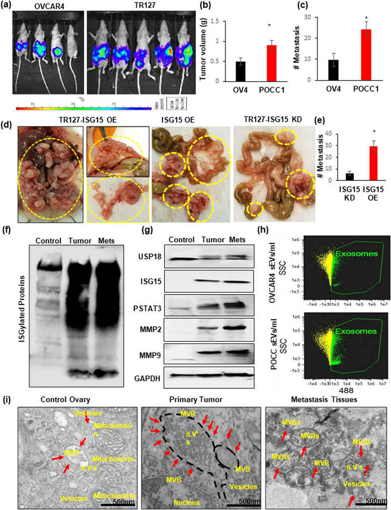
ISG15 overexpression of HGSOC ascites cells increased ovarian tumour progression and metastasis: (a) Luciferase stable transfected OVCAR4 or TR127 (1 × 10⁵ cells) were injected into the ovarian bursa in nude mice; (b and c) Tumour volume and metastasis analysed at week 5 in both groups (n = 5 mice ± SD). (d and e) HGSOC progression and metastasis observed in nude mice after intraperitoneal injections of ISG‐OE and ‐KD TR127 (2 × 10⁶ cells) (n = 4 mice ± SD). (f and g) ISG15 with ISGylation products, USP18 and downstream target proteins STAT3, MMP2 and MMP9, expression analysed in control ovary, primary and metastatic ovarian tumour tissues in mice (n = 5 mice ± SD). (h) Extracellular vesicle quantification by image stream flow cytometry in ascites developed in OVCAR4 and TR127 injected mice (n = 3 samples ± SD). (i) Increased sEV formation observed by Transmission Electron Microscopy (TEM) in primary and metastatic mice ovarian tumour tissues when compared to control ovarian tissue.
