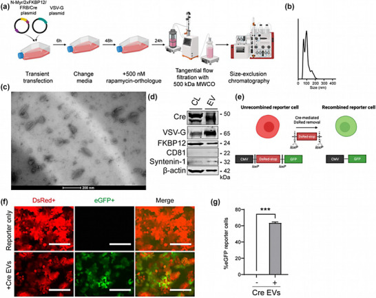
FIGURE 1.

Characterization of Cre‐EVs. (a) Schematic illustration of Cre‐EV production, the concentration of conditional media with tangential flow filtration, and purification with size‐exclusion chromatography. (b) Nanoparticle Tracking Analysis showed Cre‐EVs had a size distribution between 50–200 nm. (c) Transmission electron microscopy showed that Cre‐ EVs have a typical ‘cup‐shaped’ morphology. (d) Western blot analysis of Cre‐EVs and cell lysate of their donor cells (CL) for Cre, VSV‐G, FKBP12, CD81, Syntenin‐1, and β‐actin. (e) Schematic illustration of Cre‐mediated recombination in Cre‐loxP stoplight reporter cells. The administration of Cre‐EVs to Cre‐loxP stoplight reporter cells resulted in intracellular delivery of Cre recombinase, leading to Cre‐recombined eGFP+ reporter cells as determined by (f) microscopic and (g) flow cytometric analysis. Scale bar = 200 μm. Representative data of three individual experiments. Data are represented as mean ± SEM. Statistical analysis was performed with an unpaired t‐test, p‐values *<0.05, **<0.01, ***<0.001, and ****<0.0001 are considered statistically significant. Representative images from three individual experiments.
