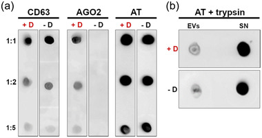
FIGURE 4.

Analysis of AT localization onto EVs. EVs from plasma samples of healthy donors were spotted on nitrocellulose membrane and analyzed with dot‐blot. All the tests were performed in presence (+D) and absence (‐D) of 0.1% (v/v) Tween‐20. Shown is a representative image of one of the three experiments performed. (a) Immunoblot vs. CD63 (EV membrane‐associated protein), Ago2 (lumen protein), and AT in presence and absence of 0.1% (v/v) Tween‐20. Membrane protein (CD63) signal is not affected by the detergent, while EV lumen protein (Ago2) is revealed only when the EV membrane is disrupted. The behaviour of AT suggests the association with EV membrane rather than its encapsulation within EV lumen. (b) Immunoblot versus AT performed on plasma EVs and supernatant after trypsin treatment and in presence (+D) or absence (‐D) of 0.1% (v/v) Tween‐20. Supernatant enriches in AT after trypsin treatment, meaning AT is directly accessible to the protease action, and further suggesting its localization on the outer leaflet of EV membrane
