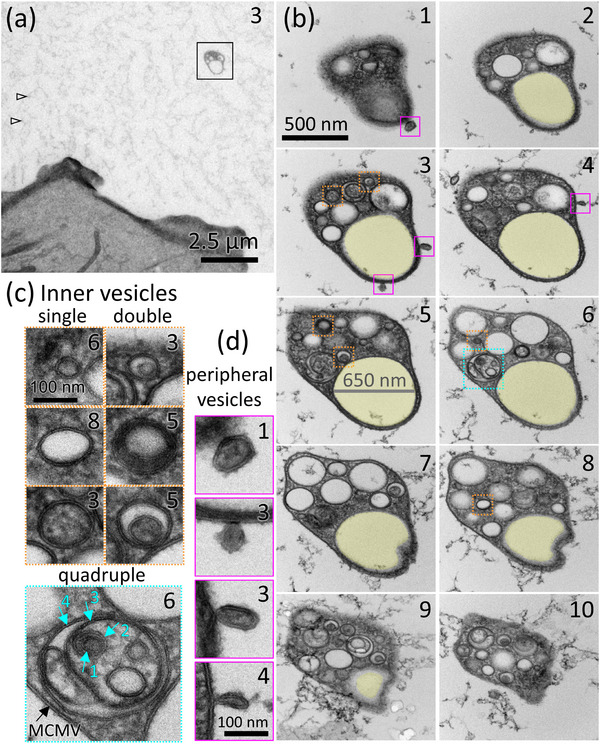
FIGURE 2.

MCMVs are found on the coverslip surface and are completely independent from neighbouring cells. (a) A low‐magnification view of a presumptive MCMV (boxed) located 7 μm beyond the periphery of the nearest cell. Arrows point to fuzzy material that is typically observed in freeze‐substituted culture medium. The complete series of serial sections through the MCMV boxed in (a) is shown in (b) and Supplemental Movie 5. The number in the upper right corner indicates the number of the serial section (ascending number indicates moving higher in the z‐axis with respect to the surface of the coverslip). Examples of vesicles inside the MCMV are boxed in orange or turquoise and shown in (c). A 650‐nm diameter MVB‐like vesicle that spans almost the entire series of sections but is devoid of ILVs is shaded yellow in (b). (c) Examples of MCMV inner vesicles that are single vesicles or double vesicles, boxed orange, or a quadruple vesicle, boxed in turquoise. Turquoise arrows indicate four layers of membrane of the quadruple vesicle, inside the MCMV limiting membrane (black arrow), amounting to five membrane layers. Inner vesicles displayed various combinations of dark, light, or granular lumens. (d) Small vesicles associated with the outer periphery of the MCMV are boxed in magenta in (b) and shown enlarged in (d).
